negative regulation of translational elongation / positive regulation of rRNA processing / nucleoid / ribosomal small subunit binding / rRNA processing / large ribosomal subunit / transferase activity / ribosome biogenesis / ribosomal small subunit biogenesis / 5S rRNA binding ...negative regulation of translational elongation / positive regulation of rRNA processing / nucleoid / ribosomal small subunit binding / rRNA processing / large ribosomal subunit / transferase activity / ribosome biogenesis / ribosomal small subunit biogenesis / 5S rRNA binding / ribosomal large subunit assembly / small ribosomal subunit / small ribosomal subunit rRNA binding / large ribosomal subunit rRNA binding / cytosolic small ribosomal subunit / cytosolic large ribosomal subunit / cytoplasmic translation / tRNA binding / negative regulation of translation / rRNA binding / structural constituent of ribosome / ribosome / translation / ribonucleoprotein complex / response to antibiotic / mRNA binding / DNA binding / RNA binding / zinc ion binding / metal ion binding / cytoplasm / cytosol Similarity search - Function
Ribosome hibernation promoting factor, long/plastid / Sigma 54 modulation/S30EA ribosomal protein, C-terminal / Sigma 54 modulation/S30EA ribosomal protein, C-terminal domain superfamily / Sigma 54 modulation/S30EA ribosomal protein C terminus / : / Ribosome hibernation promoting factor/RaiA / Ribosome hibernation promotion factor-like / Sigma 54 modulation protein / S30EA ribosomal protein / Ribosomal protein L10, eubacterial, conserved site / Ribosomal protein L10 signature. ...Ribosome hibernation promoting factor, long/plastid / Sigma 54 modulation/S30EA ribosomal protein, C-terminal / Sigma 54 modulation/S30EA ribosomal protein, C-terminal domain superfamily / Sigma 54 modulation/S30EA ribosomal protein C terminus / : / Ribosome hibernation promoting factor/RaiA / Ribosome hibernation promotion factor-like / Sigma 54 modulation protein / S30EA ribosomal protein / Ribosomal protein L10, eubacterial, conserved site / Ribosomal protein L10 signature. / Ribosomal protein L10 / : / : / Ribosomal protein S14, type Z / Ribosomal protein L31 type A / Ribosomal protein S16, conserved site / Ribosomal protein S16 signature. / Ribosomal protein L31 signature. / Ribosomal protein L31 / Ribosomal protein L31 superfamily / Ribosomal protein L31 / Ribosomal protein L10-like domain superfamily / Ribosomal protein L10P / Ribosomal protein L10 / Ribosomal protein L16 signature 1. / Ribosomal protein L6, conserved site / Ribosomal protein L6 signature 1. / Ribosomal protein L21, conserved site / Ribosomal protein L21 signature. / : / Ribosomal protein L16 signature 2. / Ribosomal protein L16, conserved site / Ribosomal protein L17 signature. / Ribosomal protein L36 signature. / : / Ribosomal protein L28/L24 superfamily / Ribosomal protein L33, conserved site / Ribosomal protein L33 signature. / Ribosomal protein L32p, bacterial type / Ribosomal protein L35, conserved site / Ribosomal protein L35 signature. / Ribosomal protein L28 / Ribosomal protein L35, non-mitochondrial / Ribosomal protein L18, bacterial-type / Ribosomal protein S6, conserved site / Ribosomal protein S6 signature. / Ribosomal protein S3, bacterial-type / Ribosomal protein S13, bacterial-type / Ribosomal protein S19, bacterial-type / : / Ribosomal protein L6, bacterial-type / Ribosomal protein S7, bacterial/organellar-type / Ribosomal protein S11, bacterial-type / Ribosomal protein S20 / Ribosomal protein S20 superfamily / Ribosomal protein S20 / Ribosomal protein S4, bacterial-type / Ribosomal protein L5, bacterial-type / Ribosomal protein S5, bacterial-type / Ribosomal protein L19, conserved site / Ribosomal protein L19 signature. / 30S ribosomal protein S17 / : / Ribosomal protein S6, plastid/chloroplast / Ribosomal protein L36 / Ribosomal protein L36 superfamily / Ribosomal protein L36 / Ribosomal protein L20 signature. / Ribosomal protein L34, conserved site / Ribosomal protein L34 signature. / Ribosomal protein S14/S29 / Ribosomal protein L14P, bacterial-type / Ribosomal protein L27, conserved site / Ribosomal protein L27 signature. / Ribosomal protein S2, bacteria/mitochondria/plastid / Ribosomal protein L35 / Ribosomal protein L35 superfamily / Ribosomal protein L22, bacterial/chloroplast-type / Ribosomal protein L35 / Ribosomal protein L2, bacterial/organellar-type / Ribosomal protein L33 / Ribosomal protein L18 / Ribosomal L18 of archaea, bacteria, mitoch. and chloroplast / Ribosomal protein S18, conserved site / Ribosomal protein S18 signature. / Ribosomal protein L33 / Ribosomal L28 family / Ribosomal protein S9, bacterial/plastid / Ribosomal protein L33 superfamily / Ribosomal protein L28/L24 / Ribosomal protein L30, bacterial-type / Ribosomal protein S16 / Ribosomal protein S16 domain superfamily / Ribosomal protein S16 / L28p-like / Ribosomal protein L16 / Ribosomal protein S15, bacterial-type / Ribosomal protein S6 / Ribosomal protein S6 / Ribosomal protein S6 superfamily Similarity search - Domain/homology
Large ribosomal subunit protein bL19 / Large ribosomal subunit protein bL32 / Small ribosomal subunit protein uS11 / Large ribosomal subunit protein bL34 / Large ribosomal subunit protein bL27 / Large ribosomal subunit protein uL24 / Large ribosomal subunit protein uL29 / Small ribosomal subunit protein uS17 / Large ribosomal subunit protein uL14 / Large ribosomal subunit protein uL5 ...Large ribosomal subunit protein bL19 / Large ribosomal subunit protein bL32 / Small ribosomal subunit protein uS11 / Large ribosomal subunit protein bL34 / Large ribosomal subunit protein bL27 / Large ribosomal subunit protein uL24 / Large ribosomal subunit protein uL29 / Small ribosomal subunit protein uS17 / Large ribosomal subunit protein uL14 / Large ribosomal subunit protein uL5 / Small ribosomal subunit protein uS14B / Small ribosomal subunit protein uS8 / Large ribosomal subunit protein uL16 / Large ribosomal subunit protein uL15 / Large ribosomal subunit protein uL30 / Large ribosomal subunit protein bL17 / Large ribosomal subunit protein bL36 / Small ribosomal subunit protein uS13 / Small ribosomal subunit protein uS2 / Small ribosomal subunit protein uS3 / Small ribosomal subunit protein uS4 / Small ribosomal subunit protein uS5 / Small ribosomal subunit protein bS6 / Small ribosomal subunit protein uS7 / Small ribosomal subunit protein uS9 / Small ribosomal subunit protein uS10 / Small ribosomal subunit protein uS12 / Small ribosomal subunit protein uS15 / Small ribosomal subunit protein bS16 / Small ribosomal subunit protein bS18 / Small ribosomal subunit protein uS19 / Small ribosomal subunit protein bS20 / Large ribosomal subunit protein bL21 / Ribosome hibernation promotion factor / Large ribosomal subunit protein bL28 / Large ribosomal subunit protein uL22 / Large ribosomal subunit protein uL2 / Large ribosomal subunit protein uL3 / Large ribosomal subunit protein uL4 / Large ribosomal subunit protein uL10 / Large ribosomal subunit protein uL23 / Large ribosomal subunit protein uL6 / Large ribosomal subunit protein uL18 / Large ribosomal subunit protein bL20 / Large ribosomal subunit protein bL35 / Large ribosomal subunit protein bL33A / Large ribosomal subunit protein uL13 / Large ribosomal subunit protein bL31 Similarity search - Component
Biological species
Bacillus subtilis subsp. subtilis str. 168 (bacteria)
Method
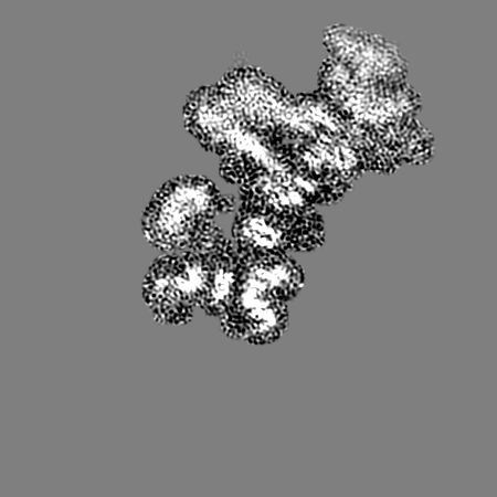
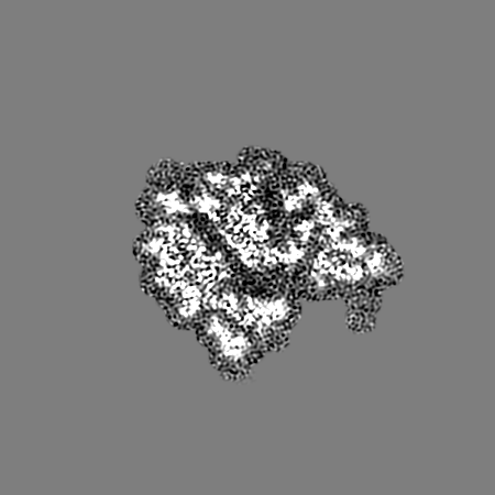
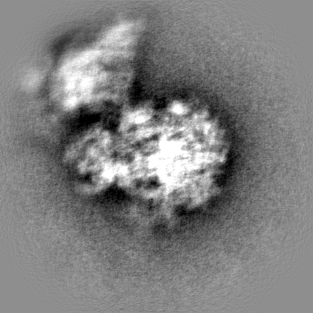
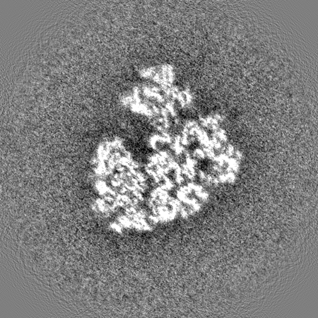
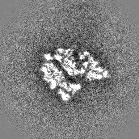
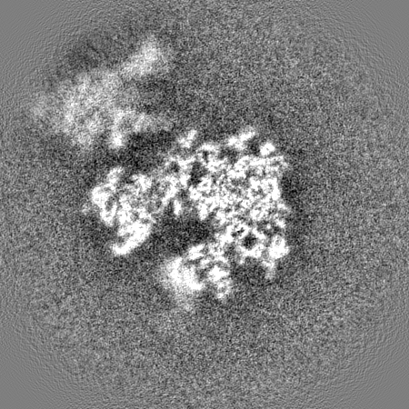
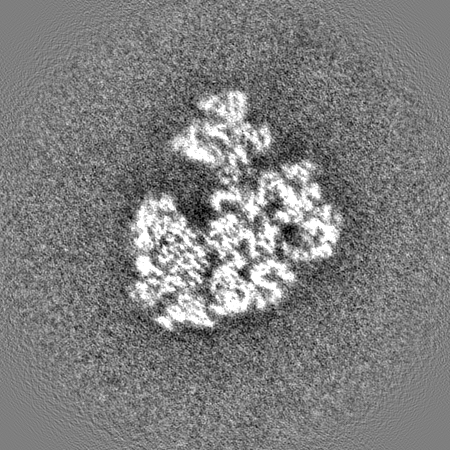
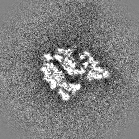
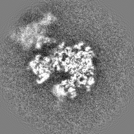
single particle reconstruction / cryo EM / Resolution: 3.8 Å
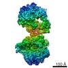
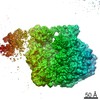
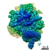
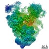
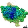
Journal: EMBO J / Year: 2017 Title: Structure of the hibernating 100S ribosome reveals the basis for 70S dimerization. Authors: Bertrand Beckert / Maha Abdelshahid / Heinrich Schäfer / Wieland Steinchen / Stefan Arenz / Otto Berninghausen / Roland Beckmann / Gert Bange / Kürşad Turgay / Daniel N Wilson / Abstract: Under stress conditions, such as nutrient deprivation, bacteria enter into a hibernation stage, which is characterized by the appearance of 100S ribosomal particles. In , dimerization of 70S ...Under stress conditions, such as nutrient deprivation, bacteria enter into a hibernation stage, which is characterized by the appearance of 100S ribosomal particles. In , dimerization of 70S ribosomes into 100S requires the action of the ribosome modulation factor (RMF) and the hibernation-promoting factor (HPF). Most other bacteria lack RMF and instead contain a long form HPF (LHPF), which is necessary and sufficient for 100S formation. While some structural information exists as to how RMF and HPF mediate formation of 100S (100S), structural insight into 100S formation by LHPF has so far been lacking. Here we present a cryo-EM structure of the hibernating 100S (100S), revealing that the C-terminal domain (CTD) of the LHPF occupies a site on the 30S platform distinct from RMF Moreover, unlike RMF, the HPF-CTD is directly involved in forming the dimer interface, thereby illustrating the divergent mechanisms by which 100S formation is mediated in the majority of bacteria that contain LHPF, compared to some γ-proteobacteria, such as .
History
Deposition
Mar 29, 2017
-
Header (metadata) release
Jun 14, 2017
-
Map release
Jun 14, 2017
-
Update
May 15, 2024
-
Current status
May 15, 2024
Processing site: PDBe / Status: Released
-
Structure visualization
Movie
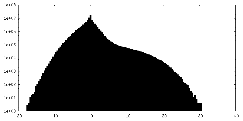
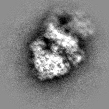
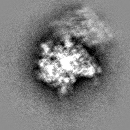
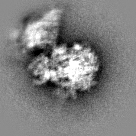
Surface view with section colored by density value
Model: Quantifoil R3/3 / Support film - topology: HOLEY / Pretreatment - Type: GLOW DISCHARGE / Pretreatment - Time: 30 sec.
Vitrification
Cryogen name: ETHANE
Details
4 OD260/ml ml Bs100S sample were applied to 2 nm pre-coated Quantifoil R3/3 holey carbon supported grids and vitrified using Vitrobot Mark IV (FEI Company)
-
Electron microscopy
Microscope
FEI TITAN KRIOS
Image recording
Film or detector model: FEI FALCON II (4k x 4k) / Detector mode: COUNTING / Average electron dose: 2.5 e/Å2
Electron beam
Acceleration voltage: 300 kV / Electron source: FIELD EMISSION GUN
Electron optics
Illumination mode: FLOOD BEAM / Imaging mode: BRIGHT FIELD / Cs: 2.7 mm
In the structure databanks used in Yorodumi, some data are registered as the other names, "COVID-19 virus" and "2019-nCoV". Here are the details of the virus and the list of structure data.
Jan 31, 2019. EMDB accession codes are about to change! (news from PDBe EMDB page)
EMDB accession codes are about to change! (news from PDBe EMDB page)
The allocation of 4 digits for EMDB accession codes will soon come to an end. Whilst these codes will remain in use, new EMDB accession codes will include an additional digit and will expand incrementally as the available range of codes is exhausted. The current 4-digit format prefixed with “EMD-” (i.e. EMD-XXXX) will advance to a 5-digit format (i.e. EMD-XXXXX), and so on. It is currently estimated that the 4-digit codes will be depleted around Spring 2019, at which point the 5-digit format will come into force.
The EM Navigator/Yorodumi systems omit the EMD- prefix.
Related info.:Q: What is EMD? / ID/Accession-code notation in Yorodumi/EM Navigator
Yorodumi is a browser for structure data from EMDB, PDB, SASBDB, etc.
This page is also the successor to EM Navigator detail page, and also detail information page/front-end page for Omokage search.
The word "yorodu" (or yorozu) is an old Japanese word meaning "ten thousand". "mi" (miru) is to see.
Related info.:EMDB / PDB / SASBDB / Comparison of 3 databanks / Yorodumi Search / Aug 31, 2016. New EM Navigator & Yorodumi / Yorodumi Papers / Jmol/JSmol / Function and homology information / Changes in new EM Navigator and Yorodumi
 Movie
Movie Controller

 Yorodumi
 Open data
 Basic information
 Map data
 Sample
 Keywords
 Function and homology information
 Authors
 Germany, 1 items
  Citation
 Structure visualization
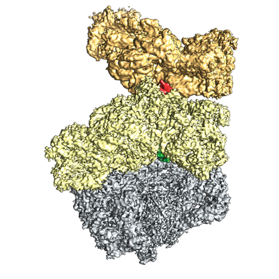
 Downloads & links
 emd_3656.png
 http://ftp.pdbj.org/pub/emdb/structures/EMD-3656


 Z (Sec.)
 Y (Row.)
 X (Col.)


 Sample components
 Processing
 Electron microscopy
 FIELD EMISSION GUN

