 Movie
Movie Controller


+ Open data

- Basic information
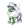
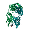
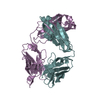
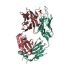
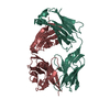
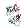
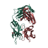
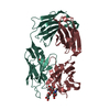
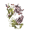
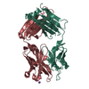
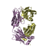
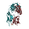
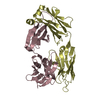
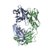
| Entry | Database: PDB / ID: 5n4g | ||||||
|---|---|---|---|---|---|---|---|
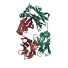
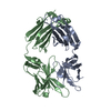
| Title | human Fab fragment 12E1 against NHBA from Neisseria meningitidis | ||||||
 Components Components |
| ||||||
 Keywords Keywords | IMMUNE SYSTEM | ||||||
| Function / homology | Immunoglobulins / Immunoglobulin-like / Sandwich / Mainly Beta Function and homology information Function and homology information | ||||||
| Biological species |  Homo sapiens (human) Homo sapiens (human) | ||||||
| Method |  X-RAY DIFFRACTION / SYNCHROTRON / MOLECULAR REPLACEMENT / Resolution: 2.75 Å X-RAY DIFFRACTION / SYNCHROTRON / MOLECULAR REPLACEMENT / Resolution: 2.75 Å | ||||||
 Authors Authors | Maritan, M. / Malito, E. | ||||||
 Citation Citation | Journal: Acta Crystallogr F Struct Biol Commun / Year: 2017 Title: Crystal structures of human Fabs targeting the Bexsero meningococcal vaccine antigen NHBA. Authors: Maritan, M. / Cozzi, R. / Lo Surdo, P. / Veggi, D. / Bottomley, M.J. / Malito, E. | ||||||
| History |
|
- Structure visualization
| Structure viewer | Molecule: MolmilJmol/JSmol |
|---|
- Downloads & links
-Download
| PDBx/mmCIF format | 5n4g.cif.gz | 100.3 KB | Display | PDBx/mmCIF format |
|---|---|---|---|---|
| PDB format | pdb5n4g.ent.gz | 74.7 KB | Display | PDB format |
| PDBx/mmJSON format | 5n4g.json.gz | Tree view | PDBx/mmJSON format | |
| Others |  Other downloads Other downloads |
-Validation report
| Arichive directory | https://data.pdbj.org/pub/pdb/validation_reports/n4/5n4gftp://data.pdbj.org/pub/pdb/validation_reports/n4/5n4g | HTTPS FTP |
|---|
-Related structure data
| Related structure data | 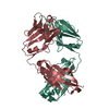 5n4jC 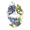 4imkS S: Starting model for refinement C: citing same article ( |
|---|---|
| Similar structure data |


-Links
 PDBj
PDBj

- Assembly
| Deposited unit | 
| ||||||||
|---|---|---|---|---|---|---|---|---|---|
| 1 |
| ||||||||
| Unit cell |
|
-Components
| #1: Antibody | Mass: 27683.893 Da / Num. of mol.: 1 Source method: isolated from a genetically manipulated source Details: fab heavy chain. the sequence provided include the cleavable tag Source: (gene. exp.) Homo sapiens (human) / Plasmid: pRS5a / Cell line (production host): HEK293 / Production host: Homo sapiens (human) | ||||||
|---|---|---|---|---|---|---|---|
| #2: Antibody | Mass: 24329.137 Da / Num. of mol.: 1 Source method: isolated from a genetically manipulated source Source: (gene. exp.) Homo sapiens (human) / Production host: Homo sapiens (human) | ||||||
| #3: Chemical |   Mass: 96.063 Da / Num. of mol.: 2 / Source method: obtained synthetically / Formula: SO4 Mass: 96.063 Da / Num. of mol.: 2 / Source method: obtained synthetically / Formula: SO4#4: Chemical | ChemComp-EDO /   Mass: 62.068 Da / Num. of mol.: 4 / Source method: obtained synthetically / Formula: C2H6O2 Mass: 62.068 Da / Num. of mol.: 4 / Source method: obtained synthetically / Formula: C2H6O2#5: Water | ChemComp-HOH / |  Mass: 18.015 Da / Num. of mol.: 73 / Source method: isolated from a natural source / Formula: H2O Mass: 18.015 Da / Num. of mol.: 73 / Source method: isolated from a natural source / Formula: H2OHas protein modification | Y | |
-Experimental details
-Experiment
| Experiment | Method: X-RAY DIFFRACTION / Number of used crystals: 1 |
|---|
- Sample preparation
| Crystal | Density Matthews: 2.59 Å3/Da / Density % sol: 52.45 % |
|---|---|
| Crystal grow | Temperature: 293.15 K / Method: vapor diffusion, sitting drop / pH: 5.6 Details: 0.2 M potassium sodium tartrate 0.1 M sodium citrate 2 M ammonium sulphate |
-Data collection
| Diffraction | Mean temperature: 100 K |
|---|---|
| Diffraction source | Source: SYNCHROTRON / Site: ESRF  / Beamline: ID23-1 / Wavelength: 0.97932 Å / Beamline: ID23-1 / Wavelength: 0.97932 Å |
| Detector | Type: ADSC QUANTUM 315r / Detector: CCD / Date: Mar 3, 2016 |
| Radiation | Protocol: SINGLE WAVELENGTH / Monochromatic (M) / Laue (L): M / Scattering type: x-ray |
| Radiation wavelength | Wavelength: 0.97932 Å / Relative weight: 1 |
| Reflection | Resolution: 2.75→45.71 Å / Num. obs: 14236 / % possible obs: 98.6 % / Redundancy: 4.6 % / Biso Wilson estimate: 68.24 Å2 / CC1/2: 0.993 / Rmerge(I) obs: 0.122 / Rpim(I) all: 0.091 / Rsym value: 0.151 / Net I/σ(I): 9.9 |
| Reflection shell | Resolution: 2.75→2.85 Å / Redundancy: 4.7 % / Rmerge(I) obs: 0.806 / Mean I/σ(I) obs: 1.6 / CC1/2: 0.609 / Rpim(I) all: 0.605 / Rsym value: 1.009 / % possible all: 97.5 |
- Processing
| Software |
| ||||||||||||||||||||||||||||||||||||||||||||||||||||||||||||||||||||||||||||||||||||||||||||||||||||||||||||||||||
|---|---|---|---|---|---|---|---|---|---|---|---|---|---|---|---|---|---|---|---|---|---|---|---|---|---|---|---|---|---|---|---|---|---|---|---|---|---|---|---|---|---|---|---|---|---|---|---|---|---|---|---|---|---|---|---|---|---|---|---|---|---|---|---|---|---|---|---|---|---|---|---|---|---|---|---|---|---|---|---|---|---|---|---|---|---|---|---|---|---|---|---|---|---|---|---|---|---|---|---|---|---|---|---|---|---|---|---|---|---|---|---|---|---|---|---|
| Refinement | Method to determine structure: MOLECULAR REPLACEMENT Starting model: 4IMK Resolution: 2.75→23.99 Å / Cor.coef. Fo:Fc: 0.892 / Cor.coef. Fo:Fc free: 0.812 / Rfactor Rfree error: 0 / SU R Cruickshank DPI: 1.404 / Cross valid method: THROUGHOUT / σ(F): 0 / SU R Blow DPI: 3.817 / SU Rfree Blow DPI: 0.362 / SU Rfree Cruickshank DPI: 0.364
| ||||||||||||||||||||||||||||||||||||||||||||||||||||||||||||||||||||||||||||||||||||||||||||||||||||||||||||||||||
| Displacement parameters | Biso mean: 56.44 Å2
| ||||||||||||||||||||||||||||||||||||||||||||||||||||||||||||||||||||||||||||||||||||||||||||||||||||||||||||||||||
| Refine analyze | Luzzati coordinate error obs: 0.36 Å | ||||||||||||||||||||||||||||||||||||||||||||||||||||||||||||||||||||||||||||||||||||||||||||||||||||||||||||||||||
| Refinement step | Cycle: 1 / Resolution: 2.75→23.99 Å
| ||||||||||||||||||||||||||||||||||||||||||||||||||||||||||||||||||||||||||||||||||||||||||||||||||||||||||||||||||
| Refine LS restraints |
| ||||||||||||||||||||||||||||||||||||||||||||||||||||||||||||||||||||||||||||||||||||||||||||||||||||||||||||||||||
| LS refinement shell | Resolution: 2.75→2.97 Å / Rfactor Rfree error: 0 / Total num. of bins used: 7
|
