 Movie
Movie Controller


+ Open data

- Basic information
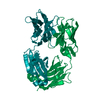
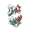
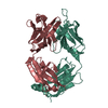
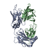
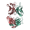
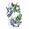
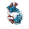
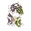
| Entry | Database: PDB / ID: 1f11 | ||||||
|---|---|---|---|---|---|---|---|
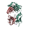
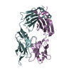
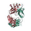
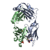
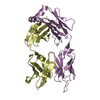
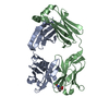
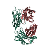
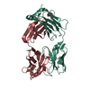
| Title | F124 FAB FRAGMENT FROM A MONOCLONAL ANTI-PRES2 ANTIBODY | ||||||
 Components Components |
| ||||||
 Keywords Keywords | IMMUNE SYSTEM / immunoglobulin / antibody / fab / hepatitis B / preS2 | ||||||
| Function / homology |  Function and homology information Function and homology informationClassical antibody-mediated complement activation / FCGR activation / Role of phospholipids in phagocytosis / Regulation of Complement cascade / positive regulation of type IIa hypersensitivity / phagocytosis, recognition / humoral immune response mediated by circulating immunoglobulin / Regulation of actin dynamics for phagocytic cup formation / positive regulation of type I hypersensitivity / antibody-dependent cellular cytotoxicity ...Classical antibody-mediated complement activation / FCGR activation / Role of phospholipids in phagocytosis / Regulation of Complement cascade / positive regulation of type IIa hypersensitivity / phagocytosis, recognition / humoral immune response mediated by circulating immunoglobulin / Regulation of actin dynamics for phagocytic cup formation / positive regulation of type I hypersensitivity / antibody-dependent cellular cytotoxicity / Fc-gamma receptor I complex binding / alpha-beta T cell receptor complex / immunoglobulin complex, circulating / phagocytosis, engulfment / IgG immunoglobulin complex / immunoglobulin receptor binding / immunoglobulin mediated immune response / complement activation, classical pathway / immunoglobulin complex / antigen binding / B cell differentiation / positive regulation of phagocytosis / positive regulation of immune response / antibacterial humoral response / adaptive immune response / defense response to bacterium / immune response / external side of plasma membrane / : / extracellular region / metal ion binding / plasma membrane / cytoplasm Similarity search - Function | ||||||
| Biological species |  | ||||||
| Method |  X-RAY DIFFRACTION / SYNCHROTRON / MOLECULAR REPLACEMENT / Resolution: 3 Å X-RAY DIFFRACTION / SYNCHROTRON / MOLECULAR REPLACEMENT / Resolution: 3 Å | ||||||
 Authors Authors | Saul, F.A. / Vulliez-Le Normand, B. / Passafiume, M. / Riottot, M.M. / Bentley, G.A. | ||||||
 Citation Citation | Journal: Acta Crystallogr.,Sect.D / Year: 2000 Title: Structure of the Fab fragment from F124, a monoclonal antibody specific for hepatitis B surface antigen. Authors: Saul, F.A. / Vulliez-Le Normand, B. / Passafiume, M. / Riottot, M.M. / Bentley, G.A. #1: Journal: FEBS Lett. / Year: 1998Title: Sequence Analysis of a Monoclonal Antibody Specific for the preS2 Region of Hepatitis B Surface Antigen, and the Cloning, Expression and Characterisation of its Single-chain Fv Construction Authors: Passafiume, M. / Vulliez-le Normand, B. / Riottot, M.M. / Bentley, G.A. | ||||||
| History |
|
- Structure visualization
| Structure viewer | Molecule: MolmilJmol/JSmol |
|---|
- Downloads & links
-Download
| PDBx/mmCIF format | 1f11.cif.gz | 172.7 KB | Display | PDBx/mmCIF format |
|---|---|---|---|---|
| PDB format | pdb1f11.ent.gz | 137.5 KB | Display | PDB format |
| PDBx/mmJSON format | 1f11.json.gz | Tree view | PDBx/mmJSON format | |
| Others |  Other downloads Other downloads |
-Validation report
| Arichive directory | https://data.pdbj.org/pub/pdb/validation_reports/f1/1f11ftp://data.pdbj.org/pub/pdb/validation_reports/f1/1f11 | HTTPS FTP |
|---|
-Related structure data
| Related structure data |  1igiS S: Starting model for refinement |
|---|---|
| Similar structure data |


-Links
 PDBj
PDBj


- Assembly
| Deposited unit | 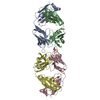
| ||||||||
|---|---|---|---|---|---|---|---|---|---|
| 1 | 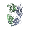
| ||||||||
| 2 | 
| ||||||||
| Unit cell |
|
-Components
| #1: Antibody | Mass: 23985.342 Da / Num. of mol.: 2 Fragment: FAB FRAGMENT (UNP P01665 residues 1-111, P01837 residues 1-106) Source method: isolated from a natural source / Source: (natural) #2: Antibody | Mass: 23594.383 Da / Num. of mol.: 2 Fragment: FAB FRAGMENT (UNP Q65ZR6 residues 18-134, P01869 residues 2-102) Source method: isolated from a natural source / Source: (natural) References: UniProt: Q65ZR6, UniProt: P01869, UniProt: Q9D8L4*PLUS Compound details | antibody F124 (isotype IgG1, kappa light chain) recognizes an epitope in the segment 120-132 of the ...antibody F124 (isotype IgG1, kappa light chain) recognizes an epitope in the segment 120-132 of the preS2 region of the hepatitis B surface antigen. | Has protein modification | Y | |
|---|
-Experimental details
-Experiment
| Experiment | Method: X-RAY DIFFRACTION / Number of used crystals: 1 |
|---|
- Sample preparation
| Crystal | Density Matthews: 2.3 Å3/Da / Density % sol: 45.73 % | ||||||||||||||||||||||||||||||||||||||||
|---|---|---|---|---|---|---|---|---|---|---|---|---|---|---|---|---|---|---|---|---|---|---|---|---|---|---|---|---|---|---|---|---|---|---|---|---|---|---|---|---|---|
| Crystal grow | Temperature: 290 K / Method: vapor diffusion, hanging drop / pH: 4.6 Details: PEG 400, sodium acetate, ammonium sulfate, pH 4.6, VAPOR DIFFUSION, HANGING DROP, temperature 290K | ||||||||||||||||||||||||||||||||||||||||
| Crystal grow | *PLUS Method: vapor diffusion | ||||||||||||||||||||||||||||||||||||||||
| Components of the solutions | *PLUS
|
-Data collection
| Diffraction | Mean temperature: 298 K |
|---|---|
| Diffraction source | Source: SYNCHROTRON / Site: LURE  / Beamline: DW32 / Wavelength: 0.96 / Beamline: DW32 / Wavelength: 0.96 |
| Detector | Type: MARRESEARCH / Detector: IMAGE PLATE / Date: Apr 1, 1999 |
| Radiation | Protocol: SINGLE WAVELENGTH / Monochromatic (M) / Laue (L): M / Scattering type: x-ray |
| Radiation wavelength | Wavelength: 0.96 Å / Relative weight: 1 |
| Reflection | Resolution: 3→30 Å / Num. all: 17261 / Num. obs: 17261 / % possible obs: 99.2 % / Observed criterion σ(F): 0 / Observed criterion σ(I): 0 / Redundancy: 1.9 % / Biso Wilson estimate: 30.6 Å2 / Rmerge(I) obs: 0.131 / Net I/σ(I): 6.7 |
| Reflection shell | Resolution: 3→3.11 Å / Redundancy: 1.9 % / Rmerge(I) obs: 0.479 / Mean I/σ(I) obs: 1.8 / Num. unique all: 1665 / % possible all: 97.5 |
| Reflection | *PLUS |
| Reflection shell | *PLUS % possible obs: 97.5 % / Num. unique obs: 1665 |
- Processing
| Software |
| ||||||||||||||||||||||||||||||||||||||||
|---|---|---|---|---|---|---|---|---|---|---|---|---|---|---|---|---|---|---|---|---|---|---|---|---|---|---|---|---|---|---|---|---|---|---|---|---|---|---|---|---|---|
| Refinement | Method to determine structure: MOLECULAR REPLACEMENT Starting model: 1IGI Resolution: 3→30 Å / σ(F): 0 / σ(I): 0 / Stereochemistry target values: Engh & Huber Details: A bulk solvent model was used. Non-crystallographic symmetry restraints were applied. NCS GROUP 1: chains A,C (1 to 109). NCS GROUP 2: chains A,C (114 to 198, 203 to 212). NCS GROUP 3: ...Details: A bulk solvent model was used. Non-crystallographic symmetry restraints were applied. NCS GROUP 1: chains A,C (1 to 109). NCS GROUP 2: chains A,C (114 to 198, 203 to 212). NCS GROUP 3: chains B,D (1 to 40, 43 to 112). NCS GROUP 4: chains B,D (115 to 127, 137 to 227).
| ||||||||||||||||||||||||||||||||||||||||
| Refinement step | Cycle: LAST / Resolution: 3→30 Å
| ||||||||||||||||||||||||||||||||||||||||
| Refine LS restraints |
| ||||||||||||||||||||||||||||||||||||||||
| Refine LS restraints NCS | NCS model details: RESTRAINED | ||||||||||||||||||||||||||||||||||||||||
| LS refinement shell | Resolution: 3→3.14 Å / Total num. of bins used: 8
| ||||||||||||||||||||||||||||||||||||||||
| Xplor file | Serial no: 1 / Param file: PARHCSDX.PRO / Topol file: TOPHCSDX.PRO | ||||||||||||||||||||||||||||||||||||||||
| Software | *PLUS Name: X-PLOR / Version: 3.851 / Classification: refinement | ||||||||||||||||||||||||||||||||||||||||
| Refinement | *PLUS Highest resolution: 3 Å / Lowest resolution: 30 Å / σ(F): 0 / % reflection Rfree: 5 % / Rfactor obs: 0.184 | ||||||||||||||||||||||||||||||||||||||||
| Solvent computation | *PLUS | ||||||||||||||||||||||||||||||||||||||||
| Displacement parameters | *PLUS Biso mean: 23.2 Å2 | ||||||||||||||||||||||||||||||||||||||||
| Refine LS restraints | *PLUS
| ||||||||||||||||||||||||||||||||||||||||
| LS refinement shell | *PLUS Rfactor Rfree: 0.35 / % reflection Rfree: 5 % / Rfactor Rwork: 0.25 / Rfactor obs: 0.271 |
