 Movie
Movie Controller

[English] 日本語
 Yorodumi
- PDB-1m7i: CRYSTAL STRUCTURE OF A MONOCLONAL FAB SPECIFIC FOR SHIGELLA FLEXN... -
+ Open data

- Basic information
| Entry | Database: PDB / ID: 1m7i | |||||||||
|---|---|---|---|---|---|---|---|---|---|---|
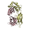
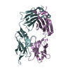
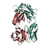
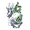
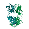
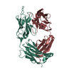
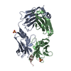
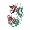
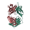
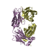
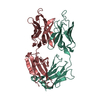
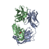
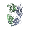
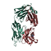
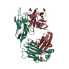
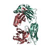
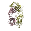
| Title | CRYSTAL STRUCTURE OF A MONOCLONAL FAB SPECIFIC FOR SHIGELLA FLEXNERI Y LIPOPOLYSACCHARIDE COMPLEXED WITH A PENTASACCHARIDE | |||||||||
 Components Components |
| |||||||||
 Keywords Keywords | IMMUNE SYSTEM / Fab-carbohydrate interactions / Shigella O-antigen / and anti-carbohydrate antibody | |||||||||
| Function / homology |  Function and homology information Function and homology informationimmunoglobulin mediated immune response / immunoglobulin complex / antigen binding / adaptive immune response / extracellular region / metal ion binding / plasma membrane Similarity search - Function | |||||||||
| Biological species |  | |||||||||
| Method |  X-RAY DIFFRACTION / SYNCHROTRON / MOLECULAR REPLACEMENT / Resolution: 2.5 Å X-RAY DIFFRACTION / SYNCHROTRON / MOLECULAR REPLACEMENT / Resolution: 2.5 Å | |||||||||
 Authors Authors | Vyas, N.K. / Vyas, M.N. / Chervenak, M.C. / Johnson, M.A. / Pinto, B.M. / Bundle, D.R. / Quiocho, F.A. | |||||||||
 Citation Citation | Journal: Biochemistry / Year: 2002 Title: Molecular Recognition of Oligosaccharide Epitopes by a Monoclonal Fab Specific for Shigella flexneri Y Lipopolysaccharide: X-ray Structures and Thermodynamics Authors: Vyas, N.K. / Vyas, M.N. / Chervenak, M.C. / Johnson, M.A. / Pinto, B.M. / Bundle, D.R. / Quiocho, F.A. #1: Journal: J.Mol.Biol. / Year: 1993Title: Preliminary Crystallographic Analysis of a Fab Specific for the O-antigen of Shigella flexneri Cell Surface Lipoplysacharide with and and without Bound Saccharides Authors: Vyas, N.K. / Vyas, M.N. / Chervenak, M.C. / Johnson, M.A. / Pinto, B.M. / Bundle, D.R. / Quiocho, F.A. | |||||||||
| History |
| |||||||||
| Remark 999 | SEQUENCE At the time of processing, this sequence of chain A and chain B have not yet been ...SEQUENCE At the time of processing, this sequence of chain A and chain B have not yet been deposited in a sequence database. |
- Structure visualization
| Structure viewer | Molecule: MolmilJmol/JSmol |
|---|
- Downloads & links
-Download
| PDBx/mmCIF format | 1m7i.cif.gz | 102.6 KB | Display | PDBx/mmCIF format |
|---|---|---|---|---|
| PDB format | pdb1m7i.ent.gz | 76.3 KB | Display | PDB format |
| PDBx/mmJSON format | 1m7i.json.gz | Tree view | PDBx/mmJSON format | |
| Others |  Other downloads Other downloads |
-Validation report
| Arichive directory | https://data.pdbj.org/pub/pdb/validation_reports/m7/1m7iftp://data.pdbj.org/pub/pdb/validation_reports/m7/1m7i | HTTPS FTP |
|---|
-Related structure data
| Related structure data |  1m71C 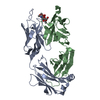 1m7dC  1mcpS S: Starting model for refinement C: citing same article ( |
|---|---|
| Similar structure data |


-Links
 PDBj
PDBj


- Assembly
| Deposited unit | 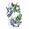
| ||||||||
|---|---|---|---|---|---|---|---|---|---|
| 1 |
| ||||||||
| Unit cell |
|
-Components
| #1: Antibody | Mass: 23717.350 Da / Num. of mol.: 1 Source method: isolated from a genetically manipulated source Source: (gene. exp.)  |
|---|---|
| #2: Antibody | Mass: 23547.361 Da / Num. of mol.: 1 Source method: isolated from a genetically manipulated source Source: (gene. exp.) |
| #3: Polysaccharide | alpha-L-rhamnopyranose-(1-2)-alpha-L-rhamnopyranose-(1-3)-alpha-L-rhamnopyranose-(1-3)-2-acetamido- ...alpha-L-rhamnopyranose-(1-2)-alpha-L-rhamnopyranose-(1-3)-alpha-L-rhamnopyranose-(1-3)-2-acetamido-2-deoxy-beta-D-glucopyranose-(1-2)-methyl 6-deoxy-alpha-L-rhamnopyranoside Source method: isolated from a genetically manipulated source |
| #4: Water | ChemComp-HOH /  Mass: 18.015 Da / Num. of mol.: 170 / Source method: isolated from a natural source / Formula: H2O Mass: 18.015 Da / Num. of mol.: 170 / Source method: isolated from a natural source / Formula: H2O |
| Has protein modification | Y |
-Experimental details
-Experiment
| Experiment | Method: X-RAY DIFFRACTION / Number of used crystals: 1 |
|---|
- Sample preparation
| Crystal | Density Matthews: 2.67 Å3/Da / Density % sol: 53.92 % |
|---|---|
| Crystal grow | Temperature: 277 K / Method: vapor diffusion / pH: 6.5 Details: MPD, potassium phosphate, pH 6.5, VAPOR DIFFUSION, temperature 277K |
| Crystal grow | *PLUS Details: Vyas, M.N., (1993) J. Mol. Biol., 231, 133. |
-Data collection
| Diffraction | Mean temperature: 281 K |
|---|---|
| Diffraction source | Source: SYNCHROTRON / Site: Photon Factory  / Beamline: BL-6A / Wavelength: 1 Å / Beamline: BL-6A / Wavelength: 1 Å |
| Detector | Type: FUJI / Detector: IMAGE PLATE / Date: Jun 2, 1990 / Details: hoton Factory BL6A2 |
| Radiation | Protocol: SINGLE WAVELENGTH / Monochromatic (M) / Laue (L): M / Scattering type: x-ray |
| Radiation wavelength | Wavelength: 1 Å / Relative weight: 1 |
| Reflection | Resolution: 2.5→20 Å / Num. all: 14764 / Num. obs: 14375 / % possible obs: 79.6 % / Observed criterion σ(I): 1.5 / Redundancy: 6.5 % / Biso Wilson estimate: 14.4 Å2 / Rmerge(I) obs: 0.097 / Rsym value: 0.075 / Net I/σ(I): 10 |
| Reflection shell | Resolution: 2.5→2.66 Å / Num. unique all: 737 / % possible all: 55.2 |
| Reflection | *PLUS Highest resolution: 2.5 Å / Num. measured all: 108148 |
| Reflection shell | *PLUS % possible obs: 55.2 % |
- Processing
| Software |
| |||||||||||||||||||||||||||
|---|---|---|---|---|---|---|---|---|---|---|---|---|---|---|---|---|---|---|---|---|---|---|---|---|---|---|---|---|
| Refinement | Method to determine structure: MOLECULAR REPLACEMENT Starting model: PDB ENTRY 1MCP Resolution: 2.5→20 Å / Rfactor Rfree error: 0.01 / Isotropic thermal model: RESTRAINED / Cross valid method: THROUGHOUT / σ(F): 0 / σ(I): 1.5 / Stereochemistry target values: Engh & Huber Details: Author states residues 127-135 of chain B is part of a disordered segment.
| |||||||||||||||||||||||||||
| Solvent computation | Solvent model: FLAT MODEL / Bsol: 32.142 Å2 / ksol: 0.302051 e/Å3 | |||||||||||||||||||||||||||
| Displacement parameters | Biso mean: 35.6 Å2
| |||||||||||||||||||||||||||
| Refine analyze |
| |||||||||||||||||||||||||||
| Refinement step | Cycle: LAST / Resolution: 2.5→20 Å
| |||||||||||||||||||||||||||
| Refine LS restraints |
| |||||||||||||||||||||||||||
| LS refinement shell | Resolution: 2.5→2.66 Å / Rfactor Rfree error: 0.048 / Total num. of bins used: 6
| |||||||||||||||||||||||||||
| Xplor file |
| |||||||||||||||||||||||||||
| Refinement | *PLUS Highest resolution: 2.5 Å / % reflection Rfree: 5 % / Rfactor Rfree: 0.279 / Rfactor Rwork: 0.207 | |||||||||||||||||||||||||||
| Solvent computation | *PLUS | |||||||||||||||||||||||||||
| Displacement parameters | *PLUS | |||||||||||||||||||||||||||
| Refine LS restraints | *PLUS
| |||||||||||||||||||||||||||
| LS refinement shell | *PLUS Rfactor Rfree: 0.411 |
