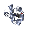
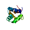
Journal: Nat Commun / Year: 2017 Title: Identification and characterization of a heterotrimeric archaeal DNA polymerase holoenzyme. Authors: Jiangyu Yan / Thomas R Beattie / Adriana L Rojas / Kelly Schermerhorn / Tamzin Gristwood / Jonathan C Trinidad / Sonja V Albers / Pietro Roversi / Andrew F Gardner / Nicola G A Abrescia / Stephen D Bell / Abstract: Since their initial characterization over 30 years ago, it has been believed that the archaeal B-family DNA polymerases are single-subunit enzymes. This contrasts with the multi-subunit B-family ...Since their initial characterization over 30 years ago, it has been believed that the archaeal B-family DNA polymerases are single-subunit enzymes. This contrasts with the multi-subunit B-family replicative polymerases of eukaryotes. Here we reveal that the highly studied PolB1 from Sulfolobus solfataricus exists as a heterotrimeric complex in cell extracts. Two small subunits, PBP1 and PBP2, associate with distinct surfaces of the larger catalytic subunit and influence the enzymatic properties of the DNA polymerase. Thus, multi-subunit replicative DNA polymerase holoenzymes are present in all three domains of life. We reveal the architecture of the assembly by a combination of cross-linking coupled with mass spectrometry, X-ray crystallography and single-particle electron microscopy. The small subunits stabilize the holoenzyme assembly and the acidic tail of one small subunit mitigates the ability of the enzyme to perform strand-displacement synthesis, with important implications for lagging strand DNA synthesis.
Mass: 18.015 Da / Num. of mol.: 30 / Source method: isolated from a natural source / Formula: H2O
Has protein modification
Y
-
Experimental details
-
Experiment
Experiment
Method: X-RAY DIFFRACTION / Number of used crystals: 1
-
Sample preparation
Crystal
Density Matthews: 2.29 Å3/Da / Density % sol: 46.28 % Preparation: FOR ANALYSIS, PLEASE, USE THE NATIVE HIGH-RESOLUTION 1.35 Ang STRUCTURE: PDB 5N41
Crystal grow
Temperature: 294.15 K / Method: vapor diffusion, sitting drop / pH: 7.5 Details: Protein: 42.7 mg/ml in 20 mM Hepes pH 7.5, 0.3 M NaCl, 1mM MgCl2 and 1mM b-ME. Crystallization buffer: 0.2 M NaNO3, 20% PEG 3350
-
Data collection
Diffraction
ID
Mean temperature (K)
Crystal-ID
1
100
1
2
100
1
3
100
1
Diffraction source
Source
Site
Beamline
ID
Wavelength (Å)
SYNCHROTRON
ESRF
ID29
1
1.71076
SYNCHROTRON
ESRF
ID29
2
1.71145
SYNCHROTRON
ESRF
ID29
3
1.70371
Detector
Type
ID
Detector
Date
DECTRIS PILATUS 6M
1
PIXEL
Nov 22, 2012
DECTRIS PILATUS 6M
2
PIXEL
Nov 22, 2012
DECTRIS PILATUS 6M
3
PIXEL
Nov 22, 2016
Radiation
ID
Protocol
Monochromatic (M) / Laue (L)
Scattering type
Wavelength-ID
1
MAD
M
x-ray
1
2
MAD
M
x-ray
2
3
MAD
M
x-ray
3
Radiation wavelength
ID
Wavelength (Å)
Relative weight
1
1.71076
1
2
1.71145
1
3
1.70371
1
Reflection
Resolution: 2.2→31.1 Å / Num. obs: 3963 / % possible obs: 99 % / Redundancy: 17 % / Biso Wilson estimate: 30.9 Å2 / Rmerge(I) obs: 0.06 / Net I/σ(I): 31.6
Reflection shell
Highest resolution: 2.2 Å / Redundancy: 8.7 % / Rmerge(I) obs: 0.07 / Mean I/σ(I) obs: 1.3 / % possible all: 90.7
-
Processing
Software
Name
Version
Classification
BUSTER
2.11.2
refinement
XDS
datareduction
SCALA
datascaling
SHARP
phasing
Refinement
Method to determine structure: MAD / Resolution: 2.24→31.08 Å / Cor.coef. Fo:Fc: 0.9153 / Cor.coef. Fo:Fc free: 0.8809 / SU R Cruickshank DPI: 0.304 / Cross valid method: FREE R-VALUE / σ(F): 0 / SU R Blow DPI: 0.317 / SU Rfree Blow DPI: 0.24 / SU Rfree Cruickshank DPI: 0.235 Details: The refinement was carried out with the data from remote 1.
Rfactor
Num. reflection
% reflection
Selection details
Rfree
0.2748
171
4.57 %
RANDOM
Rwork
0.2271
-
-
-
obs
0.2294
3740
93.62 %
-
Displacement parameters
Biso mean: 37.15 Å2
Baniso -1
Baniso -2
Baniso -3
1-
-1.6342 Å2
0 Å2
0 Å2
2-
-
-1.6342 Å2
0 Å2
3-
-
-
3.2683 Å2
Refine analyze
Luzzati coordinate error obs: 0.401 Å
Refinement step
Cycle: LAST / Resolution: 2.24→31.08 Å
Protein
Nucleic acid
Ligand
Solvent
Total
Num. atoms
468
0
24
30
522
Refine LS restraints
Refine-ID
Type
Dev ideal
Number
Restraint function
Weight
X-RAY DIFFRACTION
t_bond_d
0.01
521
HARMONIC
2
X-RAY DIFFRACTION
t_angle_deg
1.29
710
HARMONIC
2
X-RAY DIFFRACTION
t_dihedral_angle_d
195
SINUSOIDAL
2
X-RAY DIFFRACTION
t_incorr_chiral_ct
X-RAY DIFFRACTION
t_pseud_angle
X-RAY DIFFRACTION
t_trig_c_planes
15
HARMONIC
2
X-RAY DIFFRACTION
t_gen_planes
68
HARMONIC
5
X-RAY DIFFRACTION
t_it
521
HARMONIC
20
X-RAY DIFFRACTION
t_nbd
0
SEMIHARMONIC
5
X-RAY DIFFRACTION
t_omega_torsion
2.42
X-RAY DIFFRACTION
t_other_torsion
19.55
X-RAY DIFFRACTION
t_improper_torsion
X-RAY DIFFRACTION
t_chiral_improper_torsion
67
SEMIHARMONIC
5
X-RAY DIFFRACTION
t_sum_occupancies
X-RAY DIFFRACTION
t_utility_distance
X-RAY DIFFRACTION
t_utility_angle
X-RAY DIFFRACTION
t_utility_torsion
X-RAY DIFFRACTION
t_ideal_dist_contact
674
SEMIHARMONIC
4
LS refinement shell
Resolution: 2.24→2.5 Å / Total num. of bins used: 5
Rfactor
Num. reflection
% reflection
Rfree
0.3961
42
4.81 %
Rwork
0.3766
832
-
all
0.3776
874
-
obs
-
-
93.62 %
+
About Yorodumi
-
News
-
Feb 9, 2022. New format data for meta-information of EMDB entries
New format data for meta-information of EMDB entries
Version 3 of the EMDB header file is now the official format.
The previous official version 1.9 will be removed from the archive.
In the structure databanks used in Yorodumi, some data are registered as the other names, "COVID-19 virus" and "2019-nCoV". Here are the details of the virus and the list of structure data.
Jan 31, 2019. EMDB accession codes are about to change! (news from PDBe EMDB page)
EMDB accession codes are about to change! (news from PDBe EMDB page)
The allocation of 4 digits for EMDB accession codes will soon come to an end. Whilst these codes will remain in use, new EMDB accession codes will include an additional digit and will expand incrementally as the available range of codes is exhausted. The current 4-digit format prefixed with “EMD-” (i.e. EMD-XXXX) will advance to a 5-digit format (i.e. EMD-XXXXX), and so on. It is currently estimated that the 4-digit codes will be depleted around Spring 2019, at which point the 5-digit format will come into force.
The EM Navigator/Yorodumi systems omit the EMD- prefix.
Related info.:Q: What is EMD? / ID/Accession-code notation in Yorodumi/EM Navigator
Yorodumi is a browser for structure data from EMDB, PDB, SASBDB, etc.
This page is also the successor to EM Navigator detail page, and also detail information page/front-end page for Omokage search.
The word "yorodu" (or yorozu) is an old Japanese word meaning "ten thousand". "mi" (miru) is to see.
Related info.:EMDB / PDB / SASBDB / Comparison of 3 databanks / Yorodumi Search / Aug 31, 2016. New EM Navigator & Yorodumi / Yorodumi Papers / Jmol/JSmol / Function and homology information / Changes in new EM Navigator and Yorodumi
 Movie
Movie Controller


 Open data
 Basic information
 Components
 Keywords
 Function and homology information
 Sulfolobus solfataricus (archaea)
 X-RAY DIFFRACTION /
  Authors
 Spain, 1items
  Citation


 Structure visualization
 Downloads & links
 Other downloads


 PDBj
PDBj Assembly
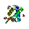


 Mass: 157.250 Da / Num. of mol.: 2 / Source method: obtained synthetically / Formula: Gd
 Mass: 92.094 Da / Num. of mol.: 3 / Source method: obtained synthetically / Formula: C3H8O3
 Mass: 62.005 Da / Num. of mol.: 1 / Source method: obtained synthetically / Formula: NO3
 Mass: 18.015 Da / Num. of mol.: 30 / Source method: isolated from a natural source / Formula: H2O
 Sample preparation
 Processing