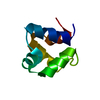
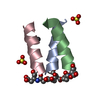
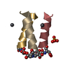
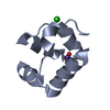
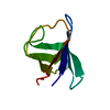
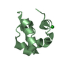
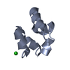
Mass: 11502.014 Da / Num. of mol.: 1 / Fragment: first SAM domain Source method: isolated from a genetically manipulated source Source: (gene. exp.) Homo sapiens (human) Description: This vector is from the NIH structural genomics consortium. It contains a His6 tag, Maltose Binding Protein tag and a TEV protease cleavage site Gene: ANKS1B / Plasmid: pDEST586 / Production host: Escherichia coli (E. coli) / Strain (production host): K12 / References: UniProt: Q7Z6G8
-
Experimental details
-
Experiment
Experiment
Method: SOLUTION NMR
NMR experiment
Conditions-ID
Experiment-ID
Solution-ID
Type
1
1
1
2D 1H-15N HSQC
1
2
1
2D 1H-13C HSQC
1
3
1
3DCBCA(CO)NH
1
4
1
3DC(CO)NH
1
5
1
3D HNCO
1
6
1
3D HN(CA)CB
1
7
1
3DH(CCO)NH
1
8
1
3D 1H-15N NOESY
1
9
1
3D 1H-13C NOESY
NMR details
Text: This structure was determined using NOE restraints and dihedral angle restraints predicted from chemical shifts
Method: torsion angle dynamics, simulated annealing / Software ordinal: 1 Details: 5000 steps using CYANA standard torsion angle dynamics, water-refinement using XPLOR-NIH and cartesian dynamics. Specifics: 2000 hot steps at 500 K, 500 cool steps to 100 K final in 100 K increments
NMR constraints
NOE constraints total: 661 / NOE intraresidue total count: 351 / NOE long range total count: 86 / NOE medium range total count: 66 / NOE sequential total count: 158
NMR representative
Selection criteria: lowest energy
NMR ensemble
Conformer selection criteria: structures with the lowest energy Conformers calculated total number: 25 / Conformers submitted total number: 10 / Maximum lower distance constraint violation: 0.5 Å / Maximum upper distance constraint violation: 0.5 Å
+
About Yorodumi
-
News
-
Feb 9, 2022. New format data for meta-information of EMDB entries
New format data for meta-information of EMDB entries
Version 3 of the EMDB header file is now the official format.
The previous official version 1.9 will be removed from the archive.
In the structure databanks used in Yorodumi, some data are registered as the other names, "COVID-19 virus" and "2019-nCoV". Here are the details of the virus and the list of structure data.
Jan 31, 2019. EMDB accession codes are about to change! (news from PDBe EMDB page)
EMDB accession codes are about to change! (news from PDBe EMDB page)
The allocation of 4 digits for EMDB accession codes will soon come to an end. Whilst these codes will remain in use, new EMDB accession codes will include an additional digit and will expand incrementally as the available range of codes is exhausted. The current 4-digit format prefixed with “EMD-” (i.e. EMD-XXXX) will advance to a 5-digit format (i.e. EMD-XXXXX), and so on. It is currently estimated that the 4-digit codes will be depleted around Spring 2019, at which point the 5-digit format will come into force.
The EM Navigator/Yorodumi systems omit the EMD- prefix.
Related info.:Q: What is EMD? / ID/Accession-code notation in Yorodumi/EM Navigator
Yorodumi is a browser for structure data from EMDB, PDB, SASBDB, etc.
This page is also the successor to EM Navigator detail page, and also detail information page/front-end page for Omokage search.
The word "yorodu" (or yorozu) is an old Japanese word meaning "ten thousand". "mi" (miru) is to see.
Related info.:EMDB / PDB / SASBDB / Comparison of 3 databanks / Yorodumi Search / Aug 31, 2016. New EM Navigator & Yorodumi / Yorodumi Papers / Jmol/JSmol / Function and homology information / Changes in new EM Navigator and Yorodumi
 Movie
Movie Controller


 Open data
 Basic information
 Components
 Keywords
 Function and homology information
 Homo sapiens (human)
 Authors
 Citation
 Structure visualization
 Downloads & links
 Other downloads
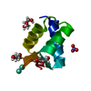


 PDBj
PDBj
 Assembly
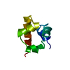

 HSQC
HSQC Sample preparation
 Processing