 Movie
Movie Controller

[English] 日本語
 Yorodumi
- PDB-5jti: Crystal structure of the human Tankyrase 1 (TNKS) SAM domain (D10... -
+ Open data

- Basic information
| Entry | Database: PDB / ID: 5jti | ||||||
|---|---|---|---|---|---|---|---|
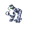
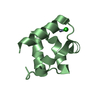
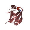
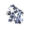
| Title | Crystal structure of the human Tankyrase 1 (TNKS) SAM domain (D1055R), crystal form 2 | ||||||
 Components Components | Tankyrase-1 | ||||||
 Keywords Keywords | SIGNALING PROTEIN / Tankyrase polymerisation Wnt signalling Poly(ADP-ribose)polymerase (PARP) / Transferase | ||||||
| Function / homology |  Function and homology information Function and homology informationnegative regulation of maintenance of mitotic sister chromatid cohesion, telomeric / positive regulation of telomere maintenance via telomere lengthening / telomerase inhibitor activity / regulation of telomere maintenance via telomerase / XAV939 stabilizes AXIN / positive regulation of telomere capping / NAD+ ADP-ribosyltransferase / protein auto-ADP-ribosylation / protein localization to chromosome, telomeric region / peptidyl-threonine phosphorylation ...negative regulation of maintenance of mitotic sister chromatid cohesion, telomeric / positive regulation of telomere maintenance via telomere lengthening / telomerase inhibitor activity / regulation of telomere maintenance via telomerase / XAV939 stabilizes AXIN / positive regulation of telomere capping / NAD+ ADP-ribosyltransferase / protein auto-ADP-ribosylation / protein localization to chromosome, telomeric region / peptidyl-threonine phosphorylation / negative regulation of telomere maintenance via telomere lengthening / NAD+-protein-aspartate ADP-ribosyltransferase activity / protein poly-ADP-ribosylation / NAD+-protein-glutamate ADP-ribosyltransferase activity / mitotic spindle pole / NAD+-protein mono-ADP-ribosyltransferase activity / pericentriolar material / Transferases; Glycosyltransferases; Pentosyltransferases / NAD+ poly-ADP-ribosyltransferase activity / nuclear pore / mRNA transport / spindle assembly / positive regulation of telomere maintenance via telomerase / nucleotidyltransferase activity / peptidyl-serine phosphorylation / mitotic spindle organization / TCF dependent signaling in response to WNT / Degradation of AXIN / Wnt signaling pathway / Regulation of PTEN stability and activity / protein polyubiquitination / positive regulation of canonical Wnt signaling pathway / protein transport / histone binding / nuclear membrane / chromosome, telomeric region / Ub-specific processing proteases / nuclear body / Golgi membrane / cell division / Golgi apparatus / positive regulation of transcription by RNA polymerase II / zinc ion binding / nucleoplasm / nucleus / cytoplasm / cytosol Similarity search - Function | ||||||
| Biological species |  Homo sapiens (human) Homo sapiens (human) | ||||||
| Method |  X-RAY DIFFRACTION / SYNCHROTRON / MOLECULAR REPLACEMENT / Resolution: 2.9 Å X-RAY DIFFRACTION / SYNCHROTRON / MOLECULAR REPLACEMENT / Resolution: 2.9 Å | ||||||
 Authors Authors | Guetter, S. / Mariotti, L. / Cronin, N. | ||||||
 Citation Citation | Journal: Mol.Cell / Year: 2016 Title: Tankyrase Requires SAM Domain-Dependent Polymerization to Support Wnt-beta-Catenin Signaling. Authors: Mariotti, L. / Templeton, C.M. / Ranes, M. / Paracuellos, P. / Cronin, N. / Beuron, F. / Morris, E. / Guettler, S. | ||||||
| History |
|
- Structure visualization
| Structure viewer | Molecule: MolmilJmol/JSmol |
|---|
- Downloads & links
-Download
| PDBx/mmCIF format | 5jti.cif.gz | 157.6 KB | Display | PDBx/mmCIF format |
|---|---|---|---|---|
| PDB format | pdb5jti.ent.gz | 126.7 KB | Display | PDB format |
| PDBx/mmJSON format | 5jti.json.gz | Tree view | PDBx/mmJSON format | |
| Others |  Other downloads Other downloads |
-Validation report
| Arichive directory | https://data.pdbj.org/pub/pdb/validation_reports/jt/5jtiftp://data.pdbj.org/pub/pdb/validation_reports/jt/5jti | HTTPS FTP |
|---|
-Related structure data
| Related structure data |  5jrtSC  5ju5C S: Starting model for refinement C: citing same article ( |
|---|---|
| Similar structure data |


-Links
 PDBj
PDBj


- Assembly
| Deposited unit | 
| ||||||||
|---|---|---|---|---|---|---|---|---|---|
| 1 | 
| ||||||||
| 2 | 
| ||||||||
| 3 | 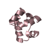
| ||||||||
| 4 | 
| ||||||||
| 5 | 
| ||||||||
| 6 | 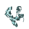
| ||||||||
| Unit cell |
|
-Components
| #1: Protein | Mass: 8854.058 Da / Num. of mol.: 6 / Fragment: UNP Residues 1018-1093 Source method: isolated from a genetically manipulated source Source: (gene. exp.) Homo sapiens (human) / Gene: TNKS, PARP5A, PARPL, TIN1, TINF1, TNKS1 / Plasmid: shuttle vector pPpT4 / Production host:  #2: Water | ChemComp-HOH / |  Mass: 18.015 Da / Num. of mol.: 62 / Source method: isolated from a natural source / Formula: H2O Mass: 18.015 Da / Num. of mol.: 62 / Source method: isolated from a natural source / Formula: H2O |
|---|
-Experimental details
-Experiment
| Experiment | Method: X-RAY DIFFRACTION / Number of used crystals: 1 |
|---|
- Sample preparation
| Crystal | Density Matthews: 3.61 Å3/Da / Density % sol: 65.9 % |
|---|---|
| Crystal grow | Temperature: 285.15 K / Method: vapor diffusion, hanging drop Details: 0.1 M Bis-Tris pH 5.5 0.2 M Magnesium Chloride 25% PEG 3350 |
-Data collection
| Diffraction | Mean temperature: 100 K |
|---|---|
| Diffraction source | Source: SYNCHROTRON / Site: Diamond  / Beamline: I03 / Wavelength: 0.9763 Å / Beamline: I03 / Wavelength: 0.9763 Å |
| Detector | Type: DECTRIS PILATUS 6M / Detector: PIXEL / Date: Aug 10, 2014 |
| Radiation | Protocol: SINGLE WAVELENGTH / Monochromatic (M) / Laue (L): M / Scattering type: x-ray |
| Radiation wavelength | Wavelength: 0.9763 Å / Relative weight: 1 |
| Reflection | Resolution: 2.9→77.41 Å / Num. obs: 15085 / % possible obs: 100 % / Redundancy: 6.6 % / Biso Wilson estimate: 77.36 Å2 / Rmerge(I) obs: 0.229 / Net I/σ(I): 7.4 |
| Reflection shell | Resolution: 2.9→3 Å |
- Processing
| Software |
| |||||||||||||||||||||||||||||||||||||||||||||||||||||||||||||||||||||||||||||||||||||||||||||||||||||||||||||||||||||||||||||||||||||||||||||||||||||||||||||||||||||||||||||||
|---|---|---|---|---|---|---|---|---|---|---|---|---|---|---|---|---|---|---|---|---|---|---|---|---|---|---|---|---|---|---|---|---|---|---|---|---|---|---|---|---|---|---|---|---|---|---|---|---|---|---|---|---|---|---|---|---|---|---|---|---|---|---|---|---|---|---|---|---|---|---|---|---|---|---|---|---|---|---|---|---|---|---|---|---|---|---|---|---|---|---|---|---|---|---|---|---|---|---|---|---|---|---|---|---|---|---|---|---|---|---|---|---|---|---|---|---|---|---|---|---|---|---|---|---|---|---|---|---|---|---|---|---|---|---|---|---|---|---|---|---|---|---|---|---|---|---|---|---|---|---|---|---|---|---|---|---|---|---|---|---|---|---|---|---|---|---|---|---|---|---|---|---|---|---|---|---|
| Refinement | Method to determine structure: MOLECULAR REPLACEMENT Starting model: 5JRT Resolution: 2.9→77.41 Å / Cor.coef. Fo:Fc: 0.93 / Cor.coef. Fo:Fc free: 0.903 / Rfactor Rfree error: 0 / SU R Cruickshank DPI: 0.694 / Cross valid method: THROUGHOUT / σ(F): 0 / SU R Blow DPI: 0.659 / SU Rfree Blow DPI: 0.304 / SU Rfree Cruickshank DPI: 0.312
| |||||||||||||||||||||||||||||||||||||||||||||||||||||||||||||||||||||||||||||||||||||||||||||||||||||||||||||||||||||||||||||||||||||||||||||||||||||||||||||||||||||||||||||||
| Displacement parameters | Biso mean: 53.1 Å2
| |||||||||||||||||||||||||||||||||||||||||||||||||||||||||||||||||||||||||||||||||||||||||||||||||||||||||||||||||||||||||||||||||||||||||||||||||||||||||||||||||||||||||||||||
| Refine analyze | Luzzati coordinate error obs: 0.37 Å | |||||||||||||||||||||||||||||||||||||||||||||||||||||||||||||||||||||||||||||||||||||||||||||||||||||||||||||||||||||||||||||||||||||||||||||||||||||||||||||||||||||||||||||||
| Refinement step | Cycle: LAST / Resolution: 2.9→77.41 Å
| |||||||||||||||||||||||||||||||||||||||||||||||||||||||||||||||||||||||||||||||||||||||||||||||||||||||||||||||||||||||||||||||||||||||||||||||||||||||||||||||||||||||||||||||
| Refine LS restraints |
| |||||||||||||||||||||||||||||||||||||||||||||||||||||||||||||||||||||||||||||||||||||||||||||||||||||||||||||||||||||||||||||||||||||||||||||||||||||||||||||||||||||||||||||||
| LS refinement shell | Resolution: 2.9→3.13 Å / Rfactor Rfree error: 0 / Total num. of bins used: 7
| |||||||||||||||||||||||||||||||||||||||||||||||||||||||||||||||||||||||||||||||||||||||||||||||||||||||||||||||||||||||||||||||||||||||||||||||||||||||||||||||||||||||||||||||
| Refinement TLS params. | Method: refined / Refine-ID: X-RAY DIFFRACTION
| |||||||||||||||||||||||||||||||||||||||||||||||||||||||||||||||||||||||||||||||||||||||||||||||||||||||||||||||||||||||||||||||||||||||||||||||||||||||||||||||||||||||||||||||
| Refinement TLS group |
|
