 Movie
Movie Controller

[English] 日本語
 Yorodumi
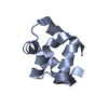
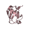
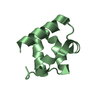
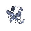
- PDB-4gu2: Crystal structure of ubiquitin from Entamoeba histolytica to 1.35... -
+ Open data

- Basic information
| Entry | Database: PDB / ID: 4gu2 | ||||||
|---|---|---|---|---|---|---|---|
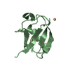
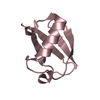
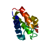
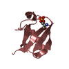
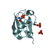
| Title | Crystal structure of ubiquitin from Entamoeba histolytica to 1.35 Angstrom | ||||||
 Components Components | Ubiquitin | ||||||
 Keywords Keywords | PROTEIN BINDING / ubiquitin / ubiquitin-like modifier / ubiquitin fold / post-translational ubiquitination / isopeptide bond / EhUbc5 / EhUba1 | ||||||
| Function / homology |  Function and homology information Function and homology informationcytosolic ribosome / modification-dependent protein catabolic process / protein tag activity / structural constituent of ribosome / protein ubiquitination / nucleus Similarity search - Function | ||||||
| Biological species |   Entamoeba histolytica (eukaryote) Entamoeba histolytica (eukaryote) | ||||||
| Method |  X-RAY DIFFRACTION / SYNCHROTRON / MOLECULAR REPLACEMENT / Resolution: 1.35 Å X-RAY DIFFRACTION / SYNCHROTRON / MOLECULAR REPLACEMENT / Resolution: 1.35 Å | ||||||
 Authors Authors | Bosch, D.E. / Siderovski, D.P. | ||||||
 Citation Citation | Journal: J.Biol.Chem. / Year: 2013 Title: Structural Determinants of Ubiquitin Conjugation in Entamoeba histolytica. Authors: Bosch, D.E. / Siderovski, D.P. | ||||||
| History |
|
- Structure visualization
| Structure viewer | Molecule: MolmilJmol/JSmol |
|---|
- Downloads & links
-Download
| PDBx/mmCIF format | 4gu2.cif.gz | 55.9 KB | Display | PDBx/mmCIF format |
|---|---|---|---|---|
| PDB format | pdb4gu2.ent.gz | 41.5 KB | Display | PDB format |
| PDBx/mmJSON format | 4gu2.json.gz | Tree view | PDBx/mmJSON format | |
| Others |  Other downloads Other downloads |
-Validation report
| Arichive directory | https://data.pdbj.org/pub/pdb/validation_reports/gu/4gu2ftp://data.pdbj.org/pub/pdb/validation_reports/gu/4gu2 | HTTPS FTP |
|---|
-Related structure data
| Related structure data |  4gprC  4gswC  1ubqS C: citing same article ( S: Starting model for refinement |
|---|---|
| Similar structure data |

-Links
 PDBj
PDBj
- Assembly
| Deposited unit | 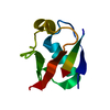
| ||||||||
|---|---|---|---|---|---|---|---|---|---|
| 1 |
| ||||||||
| Unit cell |
| ||||||||
| Components on special symmetry positions |
|
-Components
| #1: Protein | Mass: 8982.275 Da / Num. of mol.: 1 Source method: isolated from a genetically manipulated source Source: (gene. exp.) Entamoeba histolytica (eukaryote)Gene: EhUBI1, EHI_083270, EHI_083410, EHI_156660, EHI_178340 Plasmid: pLIC His / Production host:  |
|---|---|
| #2: Water | ChemComp-HOH /  Mass: 18.015 Da / Num. of mol.: 66 / Source method: isolated from a natural source / Formula: H2O Mass: 18.015 Da / Num. of mol.: 66 / Source method: isolated from a natural source / Formula: H2O |
-Experimental details
-Experiment
| Experiment | Method: X-RAY DIFFRACTION / Number of used crystals: 1 |
|---|
- Sample preparation
| Crystal | Density Matthews: 2.54 Å3/Da / Density % sol: 51.64 % |
|---|---|
| Crystal grow | Temperature: 291 K / Method: vapor diffusion, sitting drop / pH: 3.5 Details: EhUbiquitin at 17 mg/mL was mixed 1:1 with and equilibrated against crystallization solution containing 25% (w/v) PEG 3350 and 100 mM citric acid, pH 3.5, VAPOR DIFFUSION, SITTING DROP, temperature 291K |
-Data collection
| Diffraction | Mean temperature: 100 K |
|---|---|
| Diffraction source | Source: SYNCHROTRON / Site: APS  / Beamline: 23-ID-B / Wavelength: 1 / Beamline: 23-ID-B / Wavelength: 1 |
| Detector | Type: MAR scanner 300 mm plate / Detector: IMAGE PLATE / Date: Apr 8, 2012 |
| Radiation | Monochromator: double crystal / Protocol: SINGLE WAVELENGTH / Monochromatic (M) / Laue (L): M / Scattering type: x-ray |
| Radiation wavelength | Wavelength: 1 Å / Relative weight: 1 |
| Reflection | Resolution: 1.35→25.654 Å / Num. all: 20637 / Num. obs: 20493 / % possible obs: 99.3 % / Observed criterion σ(F): 0 / Observed criterion σ(I): 0 / Redundancy: 19.2 % / Biso Wilson estimate: 19.1 Å2 / Rmerge(I) obs: 0.043 / Net I/σ(I): 100 |
| Reflection shell | Resolution: 1.35→1.36 Å / Redundancy: 17.9 % / Rmerge(I) obs: 0.557 / Mean I/σ(I) obs: 6.9 / Num. unique all: 486 / % possible all: 100 |
- Processing
| Software |
| ||||||||||||||||||||||||||||||||||||||||||||||||||||||||
|---|---|---|---|---|---|---|---|---|---|---|---|---|---|---|---|---|---|---|---|---|---|---|---|---|---|---|---|---|---|---|---|---|---|---|---|---|---|---|---|---|---|---|---|---|---|---|---|---|---|---|---|---|---|---|---|---|---|
| Refinement | Method to determine structure: MOLECULAR REPLACEMENT Starting model: PDB entry 1UBQ Resolution: 1.35→25.65 Å / SU ML: 0.14 / σ(F): 1.38 / Phase error: 30.66 / Stereochemistry target values: ML
| ||||||||||||||||||||||||||||||||||||||||||||||||||||||||
| Solvent computation | Shrinkage radii: 0.9 Å / VDW probe radii: 1.11 Å / Solvent model: FLAT BULK SOLVENT MODEL | ||||||||||||||||||||||||||||||||||||||||||||||||||||||||
| Refinement step | Cycle: LAST / Resolution: 1.35→25.65 Å
| ||||||||||||||||||||||||||||||||||||||||||||||||||||||||
| Refine LS restraints |
| ||||||||||||||||||||||||||||||||||||||||||||||||||||||||
| LS refinement shell | Refine-ID: X-RAY DIFFRACTION
|
