 Movie
Movie Controller


+ Open data

- Basic information
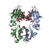
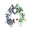
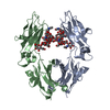
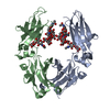
| Entry | Database: PDB / ID: 4pzn | ||||||
|---|---|---|---|---|---|---|---|
| Title | Crystal structure of PHC3 SAM L971E | ||||||
 Components Components | Polyhomeotic-like protein 3 | ||||||
 Keywords Keywords | DNA BINDING PROTEIN / SAM domain / Polycomb group / polymer / chromatin | ||||||
| Function / homology |  Function and homology information Function and homology informationPRC1 complex / PcG protein complex / SUMOylation of DNA methylation proteins / SUMOylation of RNA binding proteins / Transcriptional Regulation by E2F6 / RUNX1 interacts with co-factors whose precise effect on RUNX1 targets is not known / SUMOylation of DNA damage response and repair proteins / SUMOylation of transcription cofactors / SUMOylation of chromatin organization proteins / Regulation of PTEN gene transcription ...PRC1 complex / PcG protein complex / SUMOylation of DNA methylation proteins / SUMOylation of RNA binding proteins / Transcriptional Regulation by E2F6 / RUNX1 interacts with co-factors whose precise effect on RUNX1 targets is not known / SUMOylation of DNA damage response and repair proteins / SUMOylation of transcription cofactors / SUMOylation of chromatin organization proteins / Regulation of PTEN gene transcription / heterochromatin formation / histone binding / Oxidative Stress Induced Senescence / negative regulation of DNA-templated transcription / chromatin binding / chromatin / DNA binding / zinc ion binding / nucleoplasm / nucleus Similarity search - Function | ||||||
| Biological species |  Homo sapiens (human) Homo sapiens (human) | ||||||
| Method |  X-RAY DIFFRACTION / SYNCHROTRON / MOLECULAR REPLACEMENT / Resolution: 2.3 Å X-RAY DIFFRACTION / SYNCHROTRON / MOLECULAR REPLACEMENT / Resolution: 2.3 Å | ||||||
 Authors Authors | Nanyes, D.R. / Junco, S.E. / Taylor, A.B. / Robinson, A.K. / Patterson, N.L. / Shivarajpur, A. / Halloran, J. / Hale, S.M. / Kaur, Y. / Hart, P.J. / Kim, C.A. | ||||||
 Citation Citation | Journal: Proteins / Year: 2014 Title: Multiple polymer architectures of human polyhomeotic homolog 3 sterile alpha motif. Authors: Nanyes, D.R. / Junco, S.E. / Taylor, A.B. / Robinson, A.K. / Patterson, N.L. / Shivarajpur, A. / Halloran, J. / Hale, S.M. / Kaur, Y. / Hart, P.J. / Kim, C.A. | ||||||
| History |
|
- Structure visualization
| Structure viewer | Molecule: MolmilJmol/JSmol |
|---|
- Downloads & links
-Download
| PDBx/mmCIF format | 4pzn.cif.gz | 79.6 KB | Display | PDBx/mmCIF format |
|---|---|---|---|---|
| PDB format | pdb4pzn.ent.gz | 61 KB | Display | PDB format |
| PDBx/mmJSON format | 4pzn.json.gz | Tree view | PDBx/mmJSON format | |
| Others |  Other downloads Other downloads |
-Validation report
| Arichive directory | https://data.pdbj.org/pub/pdb/validation_reports/pz/4pznftp://data.pdbj.org/pub/pdb/validation_reports/pz/4pzn | HTTPS FTP |
|---|
-Related structure data
| Related structure data |  4pzoC 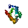 1kw4S C: citing same article ( S: Starting model for refinement |
|---|---|
| Similar structure data |


-Links
 PDBj
PDBj


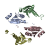
- Assembly
| Deposited unit | 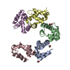
| ||||||||
|---|---|---|---|---|---|---|---|---|---|
| 1 |
| ||||||||
| 2 | 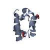
| ||||||||
| 3 | 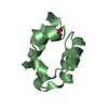
| ||||||||
| 4 | 
| ||||||||
| 5 | 
| ||||||||
| 6 | 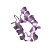
| ||||||||
| Unit cell |
|
-Components
| #1: Protein | Mass: 9465.790 Da / Num. of mol.: 5 / Fragment: sterile alpha motif / Mutation: L971E Source method: isolated from a genetically manipulated source Source: (gene. exp.) Homo sapiens (human) / Gene: EDR3, PH3, PHC3 / Plasmid: pET-3c / Production host:  #2: Chemical | ChemComp-EDO /   Mass: 62.068 Da / Num. of mol.: 5 / Source method: obtained synthetically / Formula: C2H6O2 Mass: 62.068 Da / Num. of mol.: 5 / Source method: obtained synthetically / Formula: C2H6O2#3: Water | ChemComp-HOH / |  Mass: 18.015 Da / Num. of mol.: 40 / Source method: isolated from a natural source / Formula: H2O Mass: 18.015 Da / Num. of mol.: 40 / Source method: isolated from a natural source / Formula: H2O |
|---|
-Experimental details
-Experiment
| Experiment | Method: X-RAY DIFFRACTION / Number of used crystals: 1 |
|---|
- Sample preparation
| Crystal | Density Matthews: 2.49 Å3/Da / Density % sol: 50.61 % |
|---|---|
| Crystal grow | Temperature: 295 K / Method: vapor diffusion, hanging drop / pH: 7.5 Details: 55% ethylene glycol, 100 mM tris, pH 7.5, VAPOR DIFFUSION, HANGING DROP, temperature 295K |
-Data collection
| Diffraction | Mean temperature: 100 K |
|---|---|
| Diffraction source | Source: SYNCHROTRON / Site: ALS  / Beamline: 4.2.2 / Wavelength: 1 Å / Beamline: 4.2.2 / Wavelength: 1 Å |
| Detector | Type: NOIR-1 / Detector: CCD / Date: Oct 8, 2012 |
| Radiation | Protocol: SINGLE WAVELENGTH / Monochromatic (M) / Laue (L): M / Scattering type: x-ray |
| Radiation wavelength | Wavelength: 1 Å / Relative weight: 1 |
| Reflection | Resolution: 2.3→19.68 Å / Num. obs: 19834 / % possible obs: 97.8 % / Redundancy: 2 % / Biso Wilson estimate: 44.5 Å2 / Rsym value: 0.029 / Net I/σ(I): 11.4 |
| Reflection shell | Resolution: 2.3→2.42 Å / Redundancy: 2 % / Mean I/σ(I) obs: 3.1 / Num. unique all: 2873 / Rsym value: 0.253 / % possible all: 97.2 |
- Processing
| Software |
| |||||||||||||||||||||||||||||||||||||||||||||||||||||||||||||||||||||||||||||||||||||||||||||||||||||||||
|---|---|---|---|---|---|---|---|---|---|---|---|---|---|---|---|---|---|---|---|---|---|---|---|---|---|---|---|---|---|---|---|---|---|---|---|---|---|---|---|---|---|---|---|---|---|---|---|---|---|---|---|---|---|---|---|---|---|---|---|---|---|---|---|---|---|---|---|---|---|---|---|---|---|---|---|---|---|---|---|---|---|---|---|---|---|---|---|---|---|---|---|---|---|---|---|---|---|---|---|---|---|---|---|---|---|---|
| Refinement | Method to determine structure: MOLECULAR REPLACEMENT Starting model: 1KW4 Resolution: 2.3→19.683 Å / SU ML: 0.28 / Isotropic thermal model: Isotropic / Cross valid method: THROUGHOUT / σ(F): 1.97 / Phase error: 29.52 / Stereochemistry target values: ML
| |||||||||||||||||||||||||||||||||||||||||||||||||||||||||||||||||||||||||||||||||||||||||||||||||||||||||
| Solvent computation | Shrinkage radii: 0.9 Å / VDW probe radii: 1.11 Å / Solvent model: FLAT BULK SOLVENT MODEL | |||||||||||||||||||||||||||||||||||||||||||||||||||||||||||||||||||||||||||||||||||||||||||||||||||||||||
| Displacement parameters | Biso mean: 57.6 Å2 | |||||||||||||||||||||||||||||||||||||||||||||||||||||||||||||||||||||||||||||||||||||||||||||||||||||||||
| Refinement step | Cycle: LAST / Resolution: 2.3→19.683 Å
| |||||||||||||||||||||||||||||||||||||||||||||||||||||||||||||||||||||||||||||||||||||||||||||||||||||||||
| Refine LS restraints |
| |||||||||||||||||||||||||||||||||||||||||||||||||||||||||||||||||||||||||||||||||||||||||||||||||||||||||
| LS refinement shell |
|
