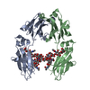
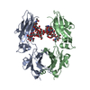
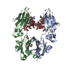
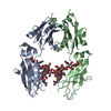
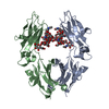
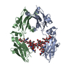
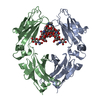
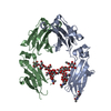
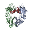
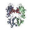
[English] 日本語
Yorodumi
- PDB-5iw6: anti-CD20 monoclonal antibody Fc fragment -

+
Open data


ID or keywords:

Loading...

-
Basic information

Entry
Database: PDB / ID: 5iw6
Titleanti-CD20 monoclonal antibody Fc fragment
Components(Ig gamma-1 chain C region) x 2
KeywordsIMMUNE SYSTEM / Glycosylation
Function / homology
Function and homology information


complement-dependent cytotoxicity / antibody-dependent cellular cytotoxicity / Fc-gamma receptor I complex binding / Classical antibody-mediated complement activation / Initial triggering of complement / immunoglobulin complex, circulating / IgG immunoglobulin complex / immunoglobulin receptor binding / FCGR activation / Role of phospholipids in phagocytosis ...complement-dependent cytotoxicity / antibody-dependent cellular cytotoxicity / Fc-gamma receptor I complex binding / Classical antibody-mediated complement activation / Initial triggering of complement / immunoglobulin complex, circulating / IgG immunoglobulin complex / immunoglobulin receptor binding / FCGR activation / Role of phospholipids in phagocytosis / complement activation, classical pathway / antigen binding / FCGR3A-mediated IL10 synthesis / Regulation of Complement cascade / FCGR3A-mediated phagocytosis / B cell receptor signaling pathway / Regulation of actin dynamics for phagocytic cup formation / antibacterial humoral response / blood microparticle / Interleukin-4 and Interleukin-13 signaling / adaptive immune response / extracellular space / extracellular exosome / extracellular region / plasma membrane
Similarity search - Function
Immunoglobulin/major histocompatibility complex, conserved site / Immunoglobulins and major histocompatibility complex proteins signature. / Immunoglobulin C-Type / Immunoglobulin C1-set / Immunoglobulin C1-set domain / Ig-like domain profile. / Immunoglobulin-like domain / Immunoglobulin-like domain superfamily / Immunoglobulins / Immunoglobulin-like fold ...Immunoglobulin/major histocompatibility complex, conserved site / Immunoglobulins and major histocompatibility complex proteins signature. / Immunoglobulin C-Type / Immunoglobulin C1-set / Immunoglobulin C1-set domain / Ig-like domain profile. / Immunoglobulin-like domain / Immunoglobulin-like domain superfamily / Immunoglobulins / Immunoglobulin-like fold / Immunoglobulin-like / Sandwich / Mainly Beta
Similarity search - Domain/homology
Immunoglobulin heavy constant gamma 1
Similarity search - Component
Biological speciesHomo sapiens (human)
MethodX-RAY DIFFRACTION / SYNCHROTRON / MOLECULAR REPLACEMENT / Resolution: 2.34 Å
AuthorsTang, C. / Chen, Z.
CitationJournal: To Be Published
Title: Structure of anti-CD20 monoclonal antibody Fc fragment at 2.34 Angstroms resolution
Authors: Tang, C. / Chen, Z.
History
DepositionMar 22, 2016Deposition site: RCSB / Processing site: PDBJ
Revision 1.0Mar 29, 2017Provider: repository / Type: Initial release
Revision 1.1Sep 27, 2017Group: Data collection / Category: diffrn_detector / Item: _diffrn_detector.detector
Revision 2.0Jul 29, 2020Group: Atomic model / Data collection ...Atomic model / Data collection / Derived calculations / Structure summary
Category: atom_site / chem_comp ...atom_site / chem_comp / entity / pdbx_branch_scheme / pdbx_chem_comp_identifier / pdbx_entity_branch / pdbx_entity_branch_descriptor / pdbx_entity_branch_link / pdbx_entity_branch_list / pdbx_entity_nonpoly / pdbx_nonpoly_scheme / pdbx_struct_assembly_gen / struct_asym / struct_conn / struct_site / struct_site_gen
Item: _atom_site.B_iso_or_equiv / _atom_site.Cartn_x ..._atom_site.B_iso_or_equiv / _atom_site.Cartn_x / _atom_site.Cartn_y / _atom_site.Cartn_z / _atom_site.auth_asym_id / _atom_site.auth_atom_id / _atom_site.auth_comp_id / _atom_site.auth_seq_id / _atom_site.label_asym_id / _atom_site.label_atom_id / _atom_site.label_comp_id / _atom_site.label_entity_id / _atom_site.type_symbol / _chem_comp.name / _chem_comp.type / _pdbx_struct_assembly_gen.asym_id_list / _struct_conn.pdbx_dist_value / _struct_conn.pdbx_role / _struct_conn.ptnr1_auth_asym_id / _struct_conn.ptnr1_auth_seq_id / _struct_conn.ptnr1_label_asym_id / _struct_conn.ptnr1_label_atom_id / _struct_conn.ptnr2_auth_asym_id / _struct_conn.ptnr2_auth_comp_id / _struct_conn.ptnr2_auth_seq_id / _struct_conn.ptnr2_label_asym_id / _struct_conn.ptnr2_label_comp_id
Description: Carbohydrate remediation / Provider: repository / Type: Remediation
Revision 2.1Nov 8, 2023Group: Data collection / Database references ...Data collection / Database references / Derived calculations / Refinement description / Structure summary
Category: chem_comp / chem_comp_atom ...chem_comp / chem_comp_atom / chem_comp_bond / database_2 / pdbx_initial_refinement_model / struct_conn
Item: _chem_comp.pdbx_synonyms / _database_2.pdbx_DOI ..._chem_comp.pdbx_synonyms / _database_2.pdbx_DOI / _database_2.pdbx_database_accession / _struct_conn.pdbx_leaving_atom_flag

-
Structure visualization

Structure viewerMolecule:
MolmilJmol/JSmol

Downloads & links

-
Assembly

Deposited unit
A: Ig gamma-1 chain C region
B: Ig gamma-1 chain C region
hetero molecules


Theoretical massNumber of molelcules
Total (without water)50,1084
Polymers47,1492
Non-polymers2,9592
Water1,928107
1


  • Idetical with deposited unit
  • defined by author&software
TypeNameSymmetry operationNumber
identity operation1_555x,y,z1
Buried area7090 Å2
ΔGint44 kcal/mol
Surface area21310 Å2
MethodPISA
Unit cell
Length a, b, c (Å)49.450, 80.004, 139.324
Angle α, β, γ (deg.)90.00, 90.00, 90.00
Int Tables number19
Space group name H-MP212121

-
Components

#1: Protein Ig gamma-1 chain C region


Mass: 23723.789 Da / Num. of mol.: 1 / Fragment: UNP residues 119-327 / Mutation: D379E, L381M
Source method: isolated from a genetically manipulated source
Source: (gene. exp.) Homo sapiens (human) / Gene: IGHG1 / Production host: Bos taurus (cattle) / References: UniProt: P01857
#2: Protein Ig gamma-1 chain C region


Mass: 23425.492 Da / Num. of mol.: 1 / Fragment: UNP residues 122-326 / Mutation: D379E, L381M
Source method: isolated from a genetically manipulated source
Source: (gene. exp.) Homo sapiens (human) / Gene: IGHG1 / Production host: Bos taurus (cattle) / References: UniProt: P01857
#3: Polysaccharide beta-D-galactopyranose-(1-4)-2-acetamido-2-deoxy-beta-D-glucopyranose-(1-2)-beta-D-mannopyranose-(1- ...beta-D-galactopyranose-(1-4)-2-acetamido-2-deoxy-beta-D-glucopyranose-(1-2)-beta-D-mannopyranose-(1-6)-[2-acetamido-2-deoxy-beta-D-glucopyranose-(1-2)-alpha-D-mannopyranose-(1-3)]beta-D-mannopyranose-(1-4)-2-acetamido-2-deoxy-beta-D-glucopyranose-(1-4)-2-acetamido-2-deoxy-beta-D-glucopyranose


Type: oligosaccharide / Mass: 1479.349 Da / Num. of mol.: 2
Source method: isolated from a genetically manipulated source
DescriptorTypeProgram
DGalpb1-4DGlcpNAcb1-2DManpb1-6[DGlcpNAcb1-2DManpa1-3]DManpb1-4DGlcpNAcb1-4DGlcpNAcb1-Glycam Condensed SequenceGMML 1.0
WURCS=2.0/4,8,7/[a2122h-1b_1-5_2*NCC/3=O][a1122h-1b_1-5][a1122h-1a_1-5][a2112h-1b_1-5]/1-1-2-3-1-2-1-4/a4-b1_b4-c1_c3-d1_c6-f1_d2-e1_f2-g1_g4-h1WURCSPDB2Glycan 1.1.0
[]{[(4+1)][b-D-GlcpNAc]{[(4+1)][b-D-GlcpNAc]{[(4+1)][b-D-Manp]{[(3+1)][a-D-Manp]{[(2+1)][b-D-GlcpNAc]{}}[(6+1)][b-D-Manp]{[(2+1)][b-D-GlcpNAc]{[(4+1)][b-D-Galp]{}}}}}}}LINUCSPDB-CARE
#4: Water ChemComp-HOH / water / Water


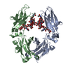
Mass: 18.015 Da / Num. of mol.: 107 / Source method: isolated from a natural source / Formula: H2O

-
Experimental details

-
Experiment

ExperimentMethod: X-RAY DIFFRACTION / Number of used crystals: 1

-
Sample preparation

CrystalDensity Matthews: 2.92 Å3/Da / Density % sol: 57.91 %
Crystal growTemperature: 291 K / Method: vapor diffusion, sitting drop / pH: 7 / Details: PEG 12000, Na HEPES

-
Data collection

DiffractionMean temperature: 80 K
Diffraction sourceSource: SYNCHROTRON / Site: SSRF / Beamline: BL18U1 / Wavelength: 1 Å
DetectorType: DECTRIS PILATUS3 6M / Detector: PIXEL / Date: Mar 21, 2015
RadiationProtocol: SINGLE WAVELENGTH / Monochromatic (M) / Laue (L): M / Scattering type: x-ray
Radiation wavelengthWavelength: 1 Å / Relative weight: 1
ReflectionResolution: 2.34→50 Å / Num. obs: 24182 / % possible obs: 99.9 % / Redundancy: 6.8 % / Rmerge(I) obs: 0.087 / Net I/σ(I): 32.1
Reflection shellResolution: 2.34→2.38 Å / Redundancy: 6.3 % / Mean I/σ(I) obs: 2.1 / % possible all: 100

-
Processing

Software
NameVersionClassification
REFMAC5.8.0049refinement
HKL-2000data reduction
HKL-2000data scaling
MOLREPphasing
RefinementMethod to determine structure: MOLECULAR REPLACEMENT
Starting model: 1L6X
Resolution: 2.34→50 Å / Cor.coef. Fo:Fc: 0.944 / Cor.coef. Fo:Fc free: 0.927 / SU B: 8.739 / SU ML: 0.2 / Cross valid method: THROUGHOUT / ESU R: 0.335 / ESU R Free: 0.241 / Stereochemistry target values: MAXIMUM LIKELIHOOD / Details: HYDROGENS HAVE BEEN ADDED IN THE RIDING POSITIONS
RfactorNum. reflection% reflectionSelection details
Rfree0.25882 1229 5.2 %RANDOM
Rwork0.22431 ---
obs0.22611 22794 99.55 %-
Solvent computationIon probe radii: 0.8 Å / Shrinkage radii: 0.8 Å / VDW probe radii: 1.2 Å / Solvent model: MASK
Displacement parametersBiso mean: 58.379 Å2
Baniso -1Baniso -2Baniso -3
1--2.73 Å20 Å2-0 Å2
2--1.61 Å2-0 Å2
3---1.12 Å2
Refinement stepCycle: LAST / Resolution: 2.34→50 Å
ProteinNucleic acidLigandSolventTotal
Num. atoms3173 0 200 107 3480
Refine LS restraints
Refine-IDTypeDev idealDev ideal targetNumber
X-RAY DIFFRACTIONr_bond_refined_d0.010.0193486
X-RAY DIFFRACTIONr_bond_other_d0.0010.023141
X-RAY DIFFRACTIONr_angle_refined_deg1.472.0234788
X-RAY DIFFRACTIONr_angle_other_deg0.77237242
X-RAY DIFFRACTIONr_dihedral_angle_1_deg6.4345405
X-RAY DIFFRACTIONr_dihedral_angle_2_deg35.81425.29138
X-RAY DIFFRACTIONr_dihedral_angle_3_deg14.80815522
X-RAY DIFFRACTIONr_dihedral_angle_4_deg9.565158
X-RAY DIFFRACTIONr_chiral_restr0.0840.2579
X-RAY DIFFRACTIONr_gen_planes_refined0.0060.0213727
X-RAY DIFFRACTIONr_gen_planes_other0.0010.02715
X-RAY DIFFRACTIONr_nbd_refined
X-RAY DIFFRACTIONr_nbd_other
X-RAY DIFFRACTIONr_nbtor_refined
X-RAY DIFFRACTIONr_nbtor_other
X-RAY DIFFRACTIONr_xyhbond_nbd_refined
X-RAY DIFFRACTIONr_xyhbond_nbd_other
X-RAY DIFFRACTIONr_metal_ion_refined
X-RAY DIFFRACTIONr_metal_ion_other
X-RAY DIFFRACTIONr_symmetry_vdw_refined
X-RAY DIFFRACTIONr_symmetry_vdw_other
X-RAY DIFFRACTIONr_symmetry_hbond_refined
X-RAY DIFFRACTIONr_symmetry_hbond_other
X-RAY DIFFRACTIONr_symmetry_metal_ion_refined
X-RAY DIFFRACTIONr_symmetry_metal_ion_other
X-RAY DIFFRACTIONr_mcbond_it3.4175.7511627
X-RAY DIFFRACTIONr_mcbond_other3.4115.751626
X-RAY DIFFRACTIONr_mcangle_it5.2958.6082028
X-RAY DIFFRACTIONr_mcangle_other5.2958.6092029
X-RAY DIFFRACTIONr_scbond_it3.3786.2961859
X-RAY DIFFRACTIONr_scbond_other3.3576.2971859
X-RAY DIFFRACTIONr_scangle_it
X-RAY DIFFRACTIONr_scangle_other5.3429.3532760
X-RAY DIFFRACTIONr_long_range_B_refined7.95846.9113734
X-RAY DIFFRACTIONr_long_range_B_other7.93746.9343709
X-RAY DIFFRACTIONr_rigid_bond_restr
X-RAY DIFFRACTIONr_sphericity_free
X-RAY DIFFRACTIONr_sphericity_bonded
LS refinement shellResolution: 2.337→2.398 Å / Total num. of bins used: 20
RfactorNum. reflection% reflection
Rfree0.329 96 -
Rwork0.319 1606 -
obs--97.65 %

+
About Yorodumi

-
News

-
Feb 9, 2022. New format data for meta-information of EMDB entries

New format data for meta-information of EMDB entries

  • Version 3 of the EMDB header file is now the official format.
  • The previous official version 1.9 will be removed from the archive.

Related info.:EMDB header

External links:wwPDB to switch to version 3 of the EMDB data model

-
Aug 12, 2020. Covid-19 info

Covid-19 info

URL: https://pdbj.org/emnavi/covid19.php

New page: Covid-19 featured information page in EM Navigator.

Related info.:Covid-19 info / Mar 5, 2020. Novel coronavirus structure data

+
Mar 5, 2020. Novel coronavirus structure data

Novel coronavirus structure data

Related info.:Yorodumi Speices / Aug 12, 2020. Covid-19 info

External links:COVID-19 featured content - PDBj / Molecule of the Month (242):Coronavirus Proteases

+
Jan 31, 2019. EMDB accession codes are about to change! (news from PDBe EMDB page)

EMDB accession codes are about to change! (news from PDBe EMDB page)

  • The allocation of 4 digits for EMDB accession codes will soon come to an end. Whilst these codes will remain in use, new EMDB accession codes will include an additional digit and will expand incrementally as the available range of codes is exhausted. The current 4-digit format prefixed with “EMD-” (i.e. EMD-XXXX) will advance to a 5-digit format (i.e. EMD-XXXXX), and so on. It is currently estimated that the 4-digit codes will be depleted around Spring 2019, at which point the 5-digit format will come into force.
  • The EM Navigator/Yorodumi systems omit the EMD- prefix.

Related info.:Q: What is EMD? / ID/Accession-code notation in Yorodumi/EM Navigator

External links:EMDB Accession Codes are Changing Soon! / Contact to PDBj

+
Jul 12, 2017. Major update of PDB

Major update of PDB

  • wwPDB released updated PDB data conforming to the new PDBx/mmCIF dictionary.
  • This is a major update changing the version number from 4 to 5, and with Remediation, in which all the entries are updated.
  • In this update, many items about electron microscopy experimental information are reorganized (e.g. em_software).
  • Now, EM Navigator and Yorodumi are based on the updated data.

External links:wwPDB Remediation / Enriched Model Files Conforming to OneDep Data Standards Now Available in the PDB FTP Archive

-
Yorodumi

Thousand views of thousand structures

  • Yorodumi is a browser for structure data from EMDB, PDB, SASBDB, etc.
  • This page is also the successor to EM Navigator detail page, and also detail information page/front-end page for Omokage search.
  • The word "yorodu" (or yorozu) is an old Japanese word meaning "ten thousand". "mi" (miru) is to see.

Related info.:EMDB / PDB / SASBDB / Comparison of 3 databanks / Yorodumi Search / Aug 31, 2016. New EM Navigator & Yorodumi / Yorodumi Papers / Jmol/JSmol / Function and homology information / Changes in new EM Navigator and Yorodumi

Read more