 Movie
Movie Controller

[English] 日本語
 Yorodumi
- PDB-1l6x: FC FRAGMENT OF RITUXIMAB BOUND TO A MINIMIZED VERSION OF THE B-DO... -
+ Open data

- Basic information
| Entry | Database: PDB / ID: 1l6x | |||||||||
|---|---|---|---|---|---|---|---|---|---|---|
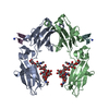
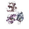
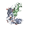
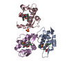
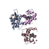
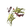
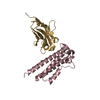
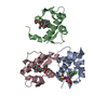
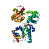
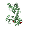
| Title | FC FRAGMENT OF RITUXIMAB BOUND TO A MINIMIZED VERSION OF THE B-DOMAIN FROM PROTEIN A CALLED Z34C | |||||||||
 Components Components |
| |||||||||
 Keywords Keywords | IMMUNE SYSTEM / IgG1 fc / protein A / fc complex / b-domain | |||||||||
| Function / homology |  Function and homology information Function and homology informationcomplement-dependent cytotoxicity / antibody-dependent cellular cytotoxicity / Fc-gamma receptor I complex binding / immunoglobulin complex, circulating / Classical antibody-mediated complement activation / immunoglobulin receptor binding / Initial triggering of complement / IgG immunoglobulin complex / FCGR activation / complement activation, classical pathway ...complement-dependent cytotoxicity / antibody-dependent cellular cytotoxicity / Fc-gamma receptor I complex binding / immunoglobulin complex, circulating / Classical antibody-mediated complement activation / immunoglobulin receptor binding / Initial triggering of complement / IgG immunoglobulin complex / FCGR activation / complement activation, classical pathway / Role of phospholipids in phagocytosis / antigen binding / FCGR3A-mediated IL10 synthesis / Regulation of Complement cascade / B cell receptor signaling pathway / FCGR3A-mediated phagocytosis / Regulation of actin dynamics for phagocytic cup formation / antibacterial humoral response / Interleukin-4 and Interleukin-13 signaling / blood microparticle / adaptive immune response / : / extracellular exosome / extracellular region / plasma membrane Similarity search - Function | |||||||||
| Biological species |  Homo sapiens (human) Homo sapiens (human) | |||||||||
| Method |  X-RAY DIFFRACTION / SYNCHROTRON / MOLECULAR REPLACEMENT / Resolution: 1.65 Å X-RAY DIFFRACTION / SYNCHROTRON / MOLECULAR REPLACEMENT / Resolution: 1.65 Å | |||||||||
 Authors Authors | Idusogie, E.E. / Presta, L.G. / Santoro-Gazzano, H. / Totpal, K. / Wong, P.Y. / Ultsch, M. / Meng, Y.G. / Mullkerrin, M.G. | |||||||||
 Citation Citation | Journal: J.Immunol. / Year: 2000 Title: Mapping of the C1q binding site on rituxan, a chimeric antibody with a human IgG1 Fc. Authors: Idusogie, E.E. / Presta, L.G. / Gazzano-Santoro, H. / Totpal, K. / Wong, P.Y. / Ultsch, M. / Meng, Y.G. / Mulkerrin, M.G. #1: Journal: J.IMMUNOL. / Year: 2001Title: Engineered Antibodies with Increased Activity to Recruit Complement Authors: Idusogie, E.E. / Wong, P.Y. / Presta, L.G. / Santoro-Gazzano, H. / Totpal, K. / Ultsch, M. / Mullkerrin, M.G. #2: Journal: MOL.CELL / Year: 2001Title: Crystal Structure at 2.8A of an FcRn/Heterodimeric Fc Complex: Mechanism of pH-Dependent Binding Authors: Martin, W.L. / West Jr., A.P. / Gan, L. / Bjorkman, P.J. #3: Journal: Proc.Natl.Acad.Sci.USA / Year: 1997Title: Structural mimicry of a native protein by a minimized binding domain Authors: Starovasnik, M.A. / Braisted, A.C. / Wells, J.A. | |||||||||
| History |
|
- Structure visualization
| Structure viewer | Molecule: MolmilJmol/JSmol |
|---|
- Downloads & links
-Download
| PDBx/mmCIF format | 1l6x.cif.gz | 78 KB | Display | PDBx/mmCIF format |
|---|---|---|---|---|
| PDB format | pdb1l6x.ent.gz | 55.5 KB | Display | PDB format |
| PDBx/mmJSON format | 1l6x.json.gz | Tree view | PDBx/mmJSON format | |
| Others |  Other downloads Other downloads |
-Validation report
| Arichive directory | https://data.pdbj.org/pub/pdb/validation_reports/l6/1l6xftp://data.pdbj.org/pub/pdb/validation_reports/l6/1l6x | HTTPS FTP |
|---|
-Related structure data
| Related structure data |  1fc1S S: Starting model for refinement |
|---|---|
| Similar structure data |

-Links
 PDBj
PDBj


- Assembly
| Deposited unit | 
| ||||||||
|---|---|---|---|---|---|---|---|---|---|
| 1 |
| ||||||||
| 2 | 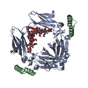
| ||||||||
| Unit cell |
|
-Components
| #1: Antibody | Mass: 23547.598 Da / Num. of mol.: 1 / Fragment: Fc Fragment, Residues 119-325 Source method: isolated from a genetically manipulated source Source: (gene. exp.) Homo sapiens (human) / Cell (production host): ovary / Production host:   Cricetulus griseus (Chinese hamster) / References: GenBank: 184747, UniProt: P01857*PLUS Cricetulus griseus (Chinese hamster) / References: GenBank: 184747, UniProt: P01857*PLUS |
|---|---|
| #2: Protein/peptide | Mass: 4190.682 Da / Num. of mol.: 1 / Fragment: ENGINEERED PEPTIDE, Residues 6-39 / Source method: obtained synthetically Details: Phage optimized sequence from the B-domain of Protein A Staphylococcus aureus. Peptide prepared using N-fluorenyl-methoxycarbonyl chemistry on Wang resin. |
| #3: Polysaccharide | beta-D-galactopyranose-(1-4)-2-acetamido-2-deoxy-beta-D-glucopyranose-(1-2)-alpha-D-mannopyranose- ...beta-D-galactopyranose-(1-4)-2-acetamido-2-deoxy-beta-D-glucopyranose-(1-2)-alpha-D-mannopyranose-(1-3)-[beta-D-galactopyranose-(1-4)-2-acetamido-2-deoxy-beta-D-glucopyranose-(1-2)-alpha-D-mannopyranose-(1-6)]beta-D-mannopyranose-(1-4)-2-acetamido-2-deoxy-beta-D-glucopyranose-(1-4)-[beta-L-fucopyranose-(1-6)]2-acetamido-2-deoxy-beta-D-glucopyranose Source method: isolated from a genetically manipulated source |
| #4: Water | ChemComp-HOH /  Mass: 18.015 Da / Num. of mol.: 360 / Source method: isolated from a natural source / Formula: H2O Mass: 18.015 Da / Num. of mol.: 360 / Source method: isolated from a natural source / Formula: H2O |
| Has protein modification | Y |
-Experimental details
-Experiment
| Experiment | Method: X-RAY DIFFRACTION / Number of used crystals: 1 |
|---|
- Sample preparation
| Crystal | Density Matthews: 2.74 Å3/Da / Density % sol: 55.04 % |
|---|---|
| Crystal grow | Temperature: 298 K / Method: vapor diffusion, sitting drop / pH: 6 Details: 20% PEG 550MME 0.1M NaOAc pH 5.5, 0.25M NaCl, pH 6.0, VAPOR DIFFUSION, SITTING DROP, temperature 298K |
-Data collection
| Diffraction | Mean temperature: 100 K |
|---|---|
| Diffraction source | Source: SYNCHROTRON / Site: SSRL  / Beamline: BL7-1 / Wavelength: 1.08 Å / Beamline: BL7-1 / Wavelength: 1.08 Å |
| Detector | Type: MARRESEARCH / Detector: IMAGE PLATE / Date: May 15, 1997 |
| Radiation | Protocol: SINGLE WAVELENGTH / Monochromatic (M) / Laue (L): M / Scattering type: x-ray |
| Radiation wavelength | Wavelength: 1.08 Å / Relative weight: 1 |
| Reflection | Resolution: 1.65→20 Å / Num. all: 35573 / Num. obs: 35573 / % possible obs: 95.4 % / Observed criterion σ(I): 0 / Redundancy: 4 % / Biso Wilson estimate: 17.4 Å2 / Rsym value: 0.036 / Net I/σ(I): 17.8 |
| Reflection shell | Resolution: 1.65→1.68 Å / Num. unique all: 1619 / Rsym value: 0.133 / % possible all: 88.7 |
- Processing
| Software |
| ||||||||||||||||||||||||||||||||||||
|---|---|---|---|---|---|---|---|---|---|---|---|---|---|---|---|---|---|---|---|---|---|---|---|---|---|---|---|---|---|---|---|---|---|---|---|---|---|
| Refinement | Method to determine structure: MOLECULAR REPLACEMENT Starting model: 1.95A Fc-Z34C complex collected at Chess. Fc was solved by molecular replacement using 2.8A 1FC1 Deisenhofer 1981 Resolution: 1.65→20 Å / Rfactor Rfree error: 0.005 / Data cutoff high absF: 10000000 / Data cutoff low absF: 0.001 / Isotropic thermal model: RESTRAINED / Cross valid method: THROUGHOUT / σ(F): 0.1 / Stereochemistry target values: Engh & Huber
| ||||||||||||||||||||||||||||||||||||
| Displacement parameters | Biso mean: 24.8 Å2
| ||||||||||||||||||||||||||||||||||||
| Refine analyze |
| ||||||||||||||||||||||||||||||||||||
| Refinement step | Cycle: LAST / Resolution: 1.65→20 Å
| ||||||||||||||||||||||||||||||||||||
| Refine LS restraints |
| ||||||||||||||||||||||||||||||||||||
| LS refinement shell | Resolution: 1.65→1.75 Å / Rfactor Rfree error: 0.013 / Total num. of bins used: 6
| ||||||||||||||||||||||||||||||||||||
| Xplor file |
|
