 Movie
Movie Controller

[English] 日本語
 Yorodumi
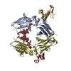
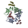
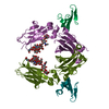
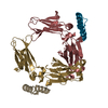
- PDB-1fc2: Crystallographic Refinement and Atomic Models of a Human FC Fragm... -
+ Open data

- Basic information
| Entry | Database: PDB / ID: 1fc2 | ||||||||||||
|---|---|---|---|---|---|---|---|---|---|---|---|---|---|
| Title | Crystallographic Refinement and Atomic Models of a Human FC Fragment and its Complex with Fragment B of Protein A from Staphylococcus Aureus at 2.9-and 2.8-Angstroms Resolution | ||||||||||||
 Components Components |
| ||||||||||||
 Keywords Keywords | IMMUNOGLOBULIN | ||||||||||||
| Function / homology |  Function and homology information Function and homology informationsymbiont-mediated suppression of host signal transduction pathway via antagonism of host cell surface receptor / complement-dependent cytotoxicity / antibody-dependent cellular cytotoxicity / Fc-gamma receptor I complex binding / IgG binding / immunoglobulin complex, circulating / Classical antibody-mediated complement activation / immunoglobulin receptor binding / Initial triggering of complement / IgG immunoglobulin complex ...symbiont-mediated suppression of host signal transduction pathway via antagonism of host cell surface receptor / complement-dependent cytotoxicity / antibody-dependent cellular cytotoxicity / Fc-gamma receptor I complex binding / IgG binding / immunoglobulin complex, circulating / Classical antibody-mediated complement activation / immunoglobulin receptor binding / Initial triggering of complement / IgG immunoglobulin complex / FCGR activation / complement activation, classical pathway / Role of phospholipids in phagocytosis / antigen binding / FCGR3A-mediated IL10 synthesis / Regulation of Complement cascade / B cell receptor signaling pathway / FCGR3A-mediated phagocytosis / Regulation of actin dynamics for phagocytic cup formation / antibacterial humoral response / Interleukin-4 and Interleukin-13 signaling / blood microparticle / adaptive immune response / : / extracellular exosome / extracellular region / plasma membrane Similarity search - Function | ||||||||||||
| Biological species |   Staphylococcus aureus subsp. aureus (bacteria) Staphylococcus aureus subsp. aureus (bacteria) Homo sapiens (human) Homo sapiens (human) | ||||||||||||
| Method |  X-RAY DIFFRACTION / Resolution: 2.8 Å X-RAY DIFFRACTION / Resolution: 2.8 Å | ||||||||||||
 Authors Authors | Deisenhofer, J. | ||||||||||||
 Citation Citation | Journal: Biochemistry / Year: 1981 Title: Crystallographic refinement and atomic models of a human Fc fragment and its complex with fragment B of protein A from Staphylococcus aureus at 2.9- and 2.8-A resolution. Authors: Deisenhofer, J. #1: Journal: Hoppe-Seyler's Z.Physiol.Chem. / Year: 1978Title: Crystallization, Crystal Structure Analysis and Atomic Model of the Complex Formed by a Human Fc Fragment and Fragment B of Protein a from Staphylococcus Aureus Authors: Deisenhofer, J. / Jones, T.A. / Huber, R. / Sjodahl, J. / Sjoquist, J. | ||||||||||||
| History |
|
- Structure visualization
| Structure viewer | Molecule: MolmilJmol/JSmol |
|---|
- Downloads & links
-Download
| PDBx/mmCIF format | 1fc2.cif.gz | 66 KB | Display | PDBx/mmCIF format |
|---|---|---|---|---|
| PDB format | pdb1fc2.ent.gz | 46 KB | Display | PDB format |
| PDBx/mmJSON format | 1fc2.json.gz | Tree view | PDBx/mmJSON format | |
| Others |  Other downloads Other downloads |
-Validation report
| Arichive directory | https://data.pdbj.org/pub/pdb/validation_reports/fc/1fc2ftp://data.pdbj.org/pub/pdb/validation_reports/fc/1fc2 | HTTPS FTP |
|---|
-Related structure data

-Links
 PDBj
PDBj


- Assembly
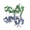
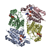
| Deposited unit | 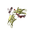
| ||||||||
|---|---|---|---|---|---|---|---|---|---|
| 1 | 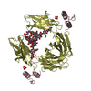
| ||||||||
| Unit cell |
| ||||||||
| Atom site foot note | 1: THESE ATOMS WERE NOT FOUND IN THE ELECTRON DENSITY MAP. THEIR COORDINATES WERE GENERATED USING STEREOCHEMICAL CRITERIA 2: RESIDUE PRO D 374 IS A CIS-PROLINE. | ||||||||
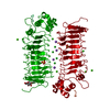
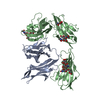
| Details | THE CRYSTALLOGRAPHIC ASYMMETRIC UNIT, FOR WHICH COORDINATES ARE PRESENTED BELOW, CONSISTS OF ONE FRAGMENT B OF PROTEIN A (CHAIN INDICATOR C BELOW) AND ONE-HALF OF AN FC FRAGMENT (CHAIN INDICATOR D BELOW) TOGETHER WITH ITS POLYSACCHARIDE. THE FOLLOWING SYMMETRY OPERATION WHEN APPLIED TO THE COORDINATES OF THE ONE-HALF OF THE FC FRAGMENT GIVEN BELOW (CHAIN D) WILL GENERATE THE SECOND HALF OF THE FC FRAGMENT MOLECULE TRNSF1 1 -0.50000 -0.86603 0.00000 0.000 TRNSF2 1 -0.86603 0.50000 0.00000 0.000 TRNSF3 1 0.00000 0.00000 -1.00000 49.133 |
-Components
| #1: Protein | Mass: 6608.254 Da / Num. of mol.: 1 Source method: isolated from a genetically manipulated source Source: (gene. exp.) Staphylococcus aureus subsp. aureus (bacteria)Strain: NCTC 8325 / References: UniProt: P02976, UniProt: P38507*PLUS |
|---|---|
| #2: Protein | Mass: 25236.615 Da / Num. of mol.: 1 Source method: isolated from a genetically manipulated source Source: (gene. exp.) Homo sapiens (human) / References: EMBL: Y14737, UniProt: P01857*PLUS |
| #3: Polysaccharide | beta-D-galactopyranose-(1-4)-2-acetamido-2-deoxy-beta-D-glucopyranose-(1-2)-alpha-D-mannopyranose- ...beta-D-galactopyranose-(1-4)-2-acetamido-2-deoxy-beta-D-glucopyranose-(1-2)-alpha-D-mannopyranose-(1-6)-[2-acetamido-2-deoxy-beta-D-glucopyranose-(1-2)-alpha-D-mannopyranose-(1-3)]alpha-D-mannopyranose-(1-4)-2-acetamido-2-deoxy-beta-D-glucopyranose-(1-4)-[alpha-L-fucopyranose-(1-6)]2-acetamido-2-deoxy-beta-D-glucopyranose Source method: isolated from a genetically manipulated source |
| #4: Chemical | ChemComp-SO4 /   Mass: 96.063 Da / Num. of mol.: 1 / Source method: obtained synthetically / Formula: SO4 Mass: 96.063 Da / Num. of mol.: 1 / Source method: obtained synthetically / Formula: SO4 |
| Has protein modification | Y |
-Experimental details
-Experiment
| Experiment | Method: X-RAY DIFFRACTION |
|---|
- Sample preparation
| Crystal | Density Matthews: 3.33 Å3/Da / Density % sol: 63.03 % | ||||||||||||||||||||||||
|---|---|---|---|---|---|---|---|---|---|---|---|---|---|---|---|---|---|---|---|---|---|---|---|---|---|
| Crystal grow | *PLUS pH: 6.8 / Method: microdialysis / Details: took Deisenhofer et al., 1976a from original paper | ||||||||||||||||||||||||
| Components of the solutions | *PLUS
|
-Data collection
| Radiation | Scattering type: x-ray |
|---|---|
| Radiation wavelength | Relative weight: 1 |
| Reflection | *PLUS Highest resolution: 2.8 Å / Num. obs: 7002 / % possible obs: 62 % / Num. measured all: 25876 |
- Processing
| Refinement | Highest resolution: 2.8 Å | ||||||||||||
|---|---|---|---|---|---|---|---|---|---|---|---|---|---|
| Refinement step | Cycle: LAST / Highest resolution: 2.8 Å
| ||||||||||||
| Refinement | *PLUS Highest resolution: 2.8 Å / Lowest resolution: 7 Å / Num. reflection all: 6243 / Rfactor Rwork: 0.24 | ||||||||||||
| Solvent computation | *PLUS | ||||||||||||
| Displacement parameters | *PLUS |
