 Movie
Movie Controller


+ Open data

- Basic information
| Entry | Database: PDB / ID: 5u4y | |||||||||
|---|---|---|---|---|---|---|---|---|---|---|
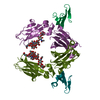
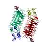
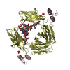
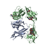
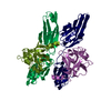
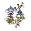
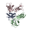
| Title | IgG Fc bound to 3 helix of the B-domain from Protein A | |||||||||
 Components Components |
| |||||||||
 Keywords Keywords | TRANSCRIPTION / IgG1 / Fc / helix / B-domain / Protein A | |||||||||
| Function / homology |  Function and homology information Function and homology informationsymbiont-mediated suppression of host signal transduction pathway via antagonism of host cell surface receptor / complement-dependent cytotoxicity / antibody-dependent cellular cytotoxicity / Fc-gamma receptor I complex binding / IgG binding / immunoglobulin complex, circulating / Classical antibody-mediated complement activation / immunoglobulin receptor binding / Initial triggering of complement / IgG immunoglobulin complex ...symbiont-mediated suppression of host signal transduction pathway via antagonism of host cell surface receptor / complement-dependent cytotoxicity / antibody-dependent cellular cytotoxicity / Fc-gamma receptor I complex binding / IgG binding / immunoglobulin complex, circulating / Classical antibody-mediated complement activation / immunoglobulin receptor binding / Initial triggering of complement / IgG immunoglobulin complex / FCGR activation / complement activation, classical pathway / Role of phospholipids in phagocytosis / immunoglobulin complex / antigen binding / FCGR3A-mediated IL10 synthesis / Regulation of Complement cascade / B cell receptor signaling pathway / FCGR3A-mediated phagocytosis / Regulation of actin dynamics for phagocytic cup formation / antibacterial humoral response / Interleukin-4 and Interleukin-13 signaling / blood microparticle / adaptive immune response / : / extracellular exosome / extracellular region / metal ion binding / plasma membrane Similarity search - Function | |||||||||
| Biological species |  Homo sapiens (human) Homo sapiens (human)  Staphylococcus aureus (bacteria) Staphylococcus aureus (bacteria) | |||||||||
| Method |  X-RAY DIFFRACTION / SYNCHROTRON / MOLECULAR REPLACEMENT / Resolution: 2.4994 Å X-RAY DIFFRACTION / SYNCHROTRON / MOLECULAR REPLACEMENT / Resolution: 2.4994 Å | |||||||||
 Authors Authors | Ultsch, M.H. / Eigenbrot, C. | |||||||||
 Citation Citation | Journal: Protein Eng. Des. Sel. / Year: 2017 Title: 3-2-1: Structural insights from stepwise shrinkage of a three-helix Fc-binding domain to a single helix. Authors: Ultsch, M. / Braisted, A. / Maun, H.R. / Eigenbrot, C. #1: Journal: Biochemistry / Year: 1981Title: Crystallographic refinement and atomic models of a human Fc fragment and its complex with fragment B of protein A from Staphylococcus aureus at 2.9- and 2.8-A resolution. Authors: Deisenhofer, J. | |||||||||
| History |
|
- Structure visualization
| Structure viewer | Molecule: MolmilJmol/JSmol |
|---|
- Downloads & links
-Download
| PDBx/mmCIF format | 5u4y.cif.gz | 237.2 KB | Display | PDBx/mmCIF format |
|---|---|---|---|---|
| PDB format | pdb5u4y.ent.gz | 191.9 KB | Display | PDB format |
| PDBx/mmJSON format | 5u4y.json.gz | Tree view | PDBx/mmJSON format | |
| Others |  Other downloads Other downloads |
-Validation report
| Arichive directory | https://data.pdbj.org/pub/pdb/validation_reports/u4/5u4yftp://data.pdbj.org/pub/pdb/validation_reports/u4/5u4y | HTTPS FTP |
|---|
-Related structure data
| Related structure data | 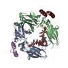 5u52C 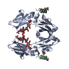 5u66C  1fc1S C: citing same article ( S: Starting model for refinement |
|---|---|
| Similar structure data |


-Links
 PDBj
PDBj


- Assembly
| Deposited unit | 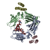
| ||||||||
|---|---|---|---|---|---|---|---|---|---|
| 1 |
| ||||||||
| Unit cell |
| ||||||||
| Components on special symmetry positions |
|
-Components
| #1: Protein | Mass: 23991.115 Da / Num. of mol.: 2 / Fragment: unp residues 261-472 Source method: isolated from a genetically manipulated source Source: (gene. exp.) Homo sapiens (human) / Gene: DKFZp686C11235 / Production host:   Cricetulus griseus (Chinese hamster) / References: UniProt: Q6MZV7, UniProt: P01857*PLUS Cricetulus griseus (Chinese hamster) / References: UniProt: Q6MZV7, UniProt: P01857*PLUS#2: Antibody | Mass: 6322.010 Da / Num. of mol.: 2 / Fragment: unp residues 101-155 Source method: isolated from a genetically manipulated source Source: (gene. exp.) Staphylococcus aureus (bacteria) / Gene: spa / Production host: #3: Polysaccharide | beta-D-galactopyranose-(1-4)-2-acetamido-2-deoxy-beta-D-glucopyranose-(1-2)-alpha-D-mannopyranose- ...beta-D-galactopyranose-(1-4)-2-acetamido-2-deoxy-beta-D-glucopyranose-(1-2)-alpha-D-mannopyranose-(1-6)-[alpha-D-mannopyranose-(1-3)]alpha-D-mannopyranose-(1-4)-2-acetamido-2-deoxy-beta-D-glucopyranose-(1-4)-[alpha-L-fucopyranose-(1-6)]2-acetamido-2-deoxy-beta-D-glucopyranose | Source method: isolated from a genetically manipulated source #4: Polysaccharide | 2-acetamido-2-deoxy-beta-D-glucopyranose-(1-2)-alpha-D-mannopyranose-(1-6)-[alpha-D-mannopyranose- ...2-acetamido-2-deoxy-beta-D-glucopyranose-(1-2)-alpha-D-mannopyranose-(1-6)-[alpha-D-mannopyranose-(1-3)]alpha-D-mannopyranose-(1-4)-2-acetamido-2-deoxy-beta-D-glucopyranose-(1-4)-[alpha-L-fucopyranose-(1-6)]2-acetamido-2-deoxy-beta-D-glucopyranose | Source method: isolated from a genetically manipulated source #5: Water | ChemComp-HOH / |  Mass: 18.015 Da / Num. of mol.: 130 / Source method: isolated from a natural source / Formula: H2O Mass: 18.015 Da / Num. of mol.: 130 / Source method: isolated from a natural source / Formula: H2OHas protein modification | Y | |
|---|
-Experimental details
-Experiment
| Experiment | Method: X-RAY DIFFRACTION / Number of used crystals: 1 |
|---|
- Sample preparation
| Crystal | Density Matthews: 2.99 Å3/Da / Density % sol: 58.92 % / Description: Rod. |
|---|---|
| Crystal grow | Temperature: 293 K / Method: vapor diffusion, sitting drop / pH: 7 Details: 25% PEG 3400, 0.1M magnesium acetate, 0.1 M HEPES pH 7.0. Protein concentration 5mg/mL. |
-Data collection
| Diffraction | Mean temperature: 273 K |
|---|---|
| Diffraction source | Source: SYNCHROTRON / Site: SSRL  / Beamline: BL7-1 / Wavelength: 1 Å / Beamline: BL7-1 / Wavelength: 1 Å |
| Detector | Type: MARRESEARCH / Detector: CCD / Date: Dec 17, 1996 |
| Radiation | Protocol: SINGLE WAVELENGTH / Monochromatic (M) / Laue (L): M / Scattering type: x-ray |
| Radiation wavelength | Wavelength: 1 Å / Relative weight: 1 |
| Reflection | Resolution: 2.4994→99 Å / Num. obs: 25214 / % possible obs: 97.1 % / Observed criterion σ(I): 2 / Redundancy: 3.2 % / Biso Wilson estimate: 43.1 Å2 / Rsym value: 0.047 / Net I/σ(I): 30.4 |
| Reflection shell | Resolution: 2.4994→2.59 Å / Redundancy: 3.2 % / Rmerge(I) obs: 0.285 / Mean I/σ(I) obs: 3.9 / % possible all: 99.1 |
- Processing
| Software |
| |||||||||||||||||||||||||||||||||||||||||||||||||||||||||||||||||||||||||||||||||||||||||||||||||||||||||||||||||||||||||||||
|---|---|---|---|---|---|---|---|---|---|---|---|---|---|---|---|---|---|---|---|---|---|---|---|---|---|---|---|---|---|---|---|---|---|---|---|---|---|---|---|---|---|---|---|---|---|---|---|---|---|---|---|---|---|---|---|---|---|---|---|---|---|---|---|---|---|---|---|---|---|---|---|---|---|---|---|---|---|---|---|---|---|---|---|---|---|---|---|---|---|---|---|---|---|---|---|---|---|---|---|---|---|---|---|---|---|---|---|---|---|---|---|---|---|---|---|---|---|---|---|---|---|---|---|---|---|---|
| Refinement | Method to determine structure: MOLECULAR REPLACEMENT Starting model: 1FC1 Resolution: 2.4994→41.075 Å / SU ML: 0.29 / Cross valid method: FREE R-VALUE / σ(F): 0.2 / Phase error: 22.52
| |||||||||||||||||||||||||||||||||||||||||||||||||||||||||||||||||||||||||||||||||||||||||||||||||||||||||||||||||||||||||||||
| Solvent computation | Shrinkage radii: 0.9 Å / VDW probe radii: 1.11 Å | |||||||||||||||||||||||||||||||||||||||||||||||||||||||||||||||||||||||||||||||||||||||||||||||||||||||||||||||||||||||||||||
| Displacement parameters | Biso mean: 53.9 Å2 | |||||||||||||||||||||||||||||||||||||||||||||||||||||||||||||||||||||||||||||||||||||||||||||||||||||||||||||||||||||||||||||
| Refinement step | Cycle: LAST / Resolution: 2.4994→41.075 Å
| |||||||||||||||||||||||||||||||||||||||||||||||||||||||||||||||||||||||||||||||||||||||||||||||||||||||||||||||||||||||||||||
| Refine LS restraints |
| |||||||||||||||||||||||||||||||||||||||||||||||||||||||||||||||||||||||||||||||||||||||||||||||||||||||||||||||||||||||||||||
| LS refinement shell |
| |||||||||||||||||||||||||||||||||||||||||||||||||||||||||||||||||||||||||||||||||||||||||||||||||||||||||||||||||||||||||||||
| Refinement TLS params. | Method: refined / Refine-ID: X-RAY DIFFRACTION
| |||||||||||||||||||||||||||||||||||||||||||||||||||||||||||||||||||||||||||||||||||||||||||||||||||||||||||||||||||||||||||||
| Refinement TLS group |
|
