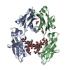
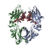
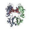
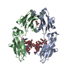
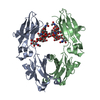
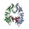
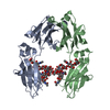
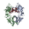
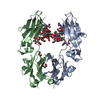
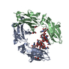
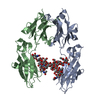
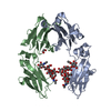
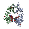
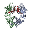
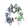
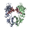
Chains A and B form the biological unit (Fc domain heavy chain contains domains CH3 and CH4)
-
Components
#1: Protein
Iggamma-1chainCregion
Mass: 24007.137 Da / Num. of mol.: 2 Fragment: Fc domain including CH2 and CH3 regions: UNP Residues 119-330 Source method: isolated from a genetically manipulated source Details: Intact, humanized IgG1 Medi-524 produced in hybridomas. Fc fragment produced by papain digestion Source: (gene. exp.) Homo sapiens (human) / Gene: IGHG1 / Production host: Mus musculus (house mouse) / Strain (production host): Mouse NSO cells / References: UniProt: P01857
In the structure databanks used in Yorodumi, some data are registered as the other names, "COVID-19 virus" and "2019-nCoV". Here are the details of the virus and the list of structure data.
Jan 31, 2019. EMDB accession codes are about to change! (news from PDBe EMDB page)
EMDB accession codes are about to change! (news from PDBe EMDB page)
The allocation of 4 digits for EMDB accession codes will soon come to an end. Whilst these codes will remain in use, new EMDB accession codes will include an additional digit and will expand incrementally as the available range of codes is exhausted. The current 4-digit format prefixed with “EMD-” (i.e. EMD-XXXX) will advance to a 5-digit format (i.e. EMD-XXXXX), and so on. It is currently estimated that the 4-digit codes will be depleted around Spring 2019, at which point the 5-digit format will come into force.
The EM Navigator/Yorodumi systems omit the EMD- prefix.
Related info.:Q: What is EMD? / ID/Accession-code notation in Yorodumi/EM Navigator
Yorodumi is a browser for structure data from EMDB, PDB, SASBDB, etc.
This page is also the successor to EM Navigator detail page, and also detail information page/front-end page for Omokage search.
The word "yorodu" (or yorozu) is an old Japanese word meaning "ten thousand". "mi" (miru) is to see.
Related info.:EMDB / PDB / SASBDB / Comparison of 3 databanks / Yorodumi Search / Aug 31, 2016. New EM Navigator & Yorodumi / Yorodumi Papers / Jmol/JSmol / Function and homology information / Changes in new EM Navigator and Yorodumi
 Movie
Movie Controller


 Open data
 Basic information
 Components
 Keywords
 Function and homology information
 Homo sapiens (human)
 X-RAY DIFFRACTION /
  Authors
 Citation
 Structure visualization
 Downloads & links
 Other downloads
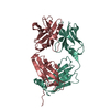

 PDBj
PDBj


 Assembly
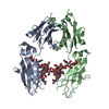

 Mass: 18.015 Da / Num. of mol.: 99 / Source method: isolated from a natural source / Formula: H2O
 Sample preparation
 Processing