 Movie
Movie Controller

[English] 日本語
 Yorodumi
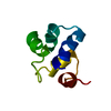
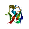
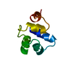
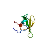
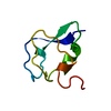
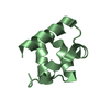
- PDB-2r63: STRUCTURAL ROLE OF A BURIED SALT BRIDGE IN THE 434 REPRESSOR DNA-... -
+ Open data

- Basic information
| Entry | Database: PDB / ID: 2r63 | ||||||
|---|---|---|---|---|---|---|---|
| Title | STRUCTURAL ROLE OF A BURIED SALT BRIDGE IN THE 434 REPRESSOR DNA-BINDING DOMAIN, NMR, 20 STRUCTURES | ||||||
 Components Components | REPRESSOR PROTEIN FROM BACTERIOPHAGE 434 | ||||||
 Keywords Keywords | GENE REGULATING PROTEIN / PHAGE 434 REPRESSOR / HELIX-TURN-HELIX / DNA-BINDING DOMAIN | ||||||
| Function / homology |  Function and homology information Function and homology information | ||||||
| Biological species |  Phage 434 (virus) Phage 434 (virus) | ||||||
| Method | SOLUTION NMR / DISTANCE GEOMETRY WITH DIANA, ENERGY MINIMIZATION WITH OPAL | ||||||
 Authors Authors | Pervushin, K.V. / Billeter, M. / Siegal, G. / Wuthrich, K. | ||||||
 Citation Citation | Journal: J.Mol.Biol. / Year: 1996 Title: Structural role of a buried salt bridge in the 434 repressor DNA-binding domain. Authors: Pervushin, K. / Billeter, M. / Siegal, G. / Wuthrich, K. #1: Journal: J.Mol.Biol. / Year: 1992Title: Determination of the Nuclear Magnetic Resonance Solution Structure of the DNA-Binding Domain (Residues 1 to 69) of the 434 Repressor and Comparison with the X-Ray Crystal Structure Authors: Neri, D. / Billeter, M. / Wuthrich, K. #2: Journal: J.Mol.Biol. / Year: 1989Title: Structure of the Amino-Terminal Domain of Phage 434 Repressor at 2.0 A Resolution Authors: Mondragon, A. / Subbiah, S. / Almo, S.C. / Drottar, M. / Harrison, S.C. | ||||||
| History |
|
- Structure visualization
| Structure viewer | Molecule: MolmilJmol/JSmol |
|---|
- Downloads & links
-Download
| PDBx/mmCIF format | 2r63.cif.gz | 423.8 KB | Display | PDBx/mmCIF format |
|---|---|---|---|---|
| PDB format | pdb2r63.ent.gz | 361.1 KB | Display | PDB format |
| PDBx/mmJSON format | 2r63.json.gz | Tree view | PDBx/mmJSON format | |
| Others |  Other downloads Other downloads |
-Validation report
| Arichive directory | https://data.pdbj.org/pub/pdb/validation_reports/r6/2r63ftp://data.pdbj.org/pub/pdb/validation_reports/r6/2r63 | HTTPS FTP |
|---|
-Related structure data


-Links
 PDBj
PDBj


- Assembly
| Deposited unit | 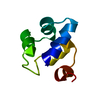
| |||||||||
|---|---|---|---|---|---|---|---|---|---|---|
| 1 |
| |||||||||
| NMR ensembles |
|
-Components
| #1: Protein | Mass: 6874.874 Da / Num. of mol.: 1 / Fragment: DNA-BINDING DOMAIN, RESIDUES 1 - 63 / Mutation: R10M Source method: isolated from a genetically manipulated source Source: (gene. exp.) Phage 434 (virus) / Genus: Lambda-like viruses / Species: Enterobacteria phage lambda / Production host:  |
|---|
-Experimental details
-Experiment
| Experiment | Method: SOLUTION NMR | ||||||||||||
|---|---|---|---|---|---|---|---|---|---|---|---|---|---|
| NMR experiment |
|
- Sample preparation
| Sample conditions | pH: 4.8 / Temperature: 286 K |
|---|---|
| Crystal grow | *PLUS Method: other / Details: NMR |
-NMR measurement
| NMR spectrometer | Type: Varian UNITY PLUS 750 / Manufacturer: Varian / Model: UNITY PLUS 750 / Field strength: 750 MHz |
|---|
- Processing
| NMR software |
| ||||||||||||||||||||
|---|---|---|---|---|---|---|---|---|---|---|---|---|---|---|---|---|---|---|---|---|---|
| Refinement | Method: DISTANCE GEOMETRY WITH DIANA, ENERGY MINIMIZATION WITH OPAL Software ordinal: 1 | ||||||||||||||||||||
| NMR ensemble | Conformer selection criteria: DIANA PENALTY FUNCTION / Conformers calculated total number: 20 / Conformers submitted total number: 20 |
