 Movie
Movie Controller


+ Open data

- Basic information
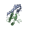
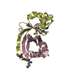
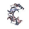
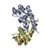
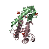
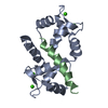
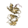
| Entry | Database: PDB / ID: 5m33 | ||||||||||||
|---|---|---|---|---|---|---|---|---|---|---|---|---|---|
| Title | Structural tuning of CD81LEL (space group P21) | ||||||||||||
 Components Components | CD81 antigen | ||||||||||||
 Keywords Keywords | CELL ADHESION / human cellular receptor for Hepatitis C virus / signaling protein | ||||||||||||
| Function / homology |  Function and homology information Function and homology informationpositive regulation of adaptive immune memory response / positive regulation of protein catabolic process in the vacuole / CD4-positive, alpha-beta T cell costimulation / positive regulation of B cell receptor signaling pathway / osteoclast fusion / myoblast fusion involved in skeletal muscle regeneration / positive regulation of T cell activation via T cell receptor contact with antigen bound to MHC molecule on antigen presenting cell / positive regulation of inflammatory response to antigenic stimulus / regulation of macrophage migration / macrophage fusion ...positive regulation of adaptive immune memory response / positive regulation of protein catabolic process in the vacuole / CD4-positive, alpha-beta T cell costimulation / positive regulation of B cell receptor signaling pathway / osteoclast fusion / myoblast fusion involved in skeletal muscle regeneration / positive regulation of T cell activation via T cell receptor contact with antigen bound to MHC molecule on antigen presenting cell / positive regulation of inflammatory response to antigenic stimulus / regulation of macrophage migration / macrophage fusion / immunological synapse formation / tetraspanin-enriched microdomain / positive regulation of T-helper 2 cell cytokine production / transferrin receptor binding / humoral immune response mediated by circulating immunoglobulin / positive regulation of protein exit from endoplasmic reticulum / protein localization to lysosome / MHC class II protein binding / positive regulation of CD4-positive, alpha-beta T cell proliferation / positive regulation of T cell receptor signaling pathway / cholesterol binding / immunological synapse / cellular response to low-density lipoprotein particle stimulus / positive regulation of receptor clustering / positive regulation of B cell proliferation / Regulation of Complement cascade / basal plasma membrane / protein localization to plasma membrane / regulation of protein stability / receptor internalization / integrin binding / Immunoregulatory interactions between a Lymphoid and a non-Lymphoid cell / MHC class II protein complex binding / virus receptor activity / vesicle / basolateral plasma membrane / positive regulation of MAPK cascade / focal adhesion / positive regulation of transcription by RNA polymerase II / extracellular exosome / membrane / plasma membrane / cytosol Similarity search - Function | ||||||||||||
| Biological species |  Homo sapiens (human) Homo sapiens (human) | ||||||||||||
| Method |  X-RAY DIFFRACTION / SYNCHROTRON / MOLECULAR REPLACEMENT / Resolution: 1.28 Å X-RAY DIFFRACTION / SYNCHROTRON / MOLECULAR REPLACEMENT / Resolution: 1.28 Å | ||||||||||||
 Authors Authors | Cunha, E.S. / Sfriso, P. / Rojas, A.L. / Roversi, P. / Hospital, A. / Orozco, M. / Abrescia, N.G. | ||||||||||||
| Funding support |  Spain, 3items Spain, 3items
| ||||||||||||
 Citation Citation | Journal: Structure / Year: 2017 Title: Mechanism of Structural Tuning of the Hepatitis C Virus Human Cellular Receptor CD81 Large Extracellular Loop. Authors: Cunha, E.S. / Sfriso, P. / Rojas, A.L. / Roversi, P. / Hospital, A. / Orozco, M. / Abrescia, N.G. | ||||||||||||
| History |
|
- Structure visualization
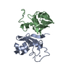
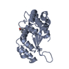
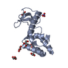
| Structure viewer | Molecule: MolmilJmol/JSmol |
|---|
- Downloads & links
-Download
| PDBx/mmCIF format | 5m33.cif.gz | 124.3 KB | Display | PDBx/mmCIF format |
|---|---|---|---|---|
| PDB format | pdb5m33.ent.gz | 99.2 KB | Display | PDB format |
| PDBx/mmJSON format | 5m33.json.gz | Tree view | PDBx/mmJSON format | |
| Others |  Other downloads Other downloads |
-Validation report
| Arichive directory | https://data.pdbj.org/pub/pdb/validation_reports/m3/5m33ftp://data.pdbj.org/pub/pdb/validation_reports/m3/5m33 | HTTPS FTP |
|---|
-Related structure data
| Related structure data | 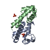 5m2cC  5m3dC  5m3tC 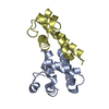 5m4rC 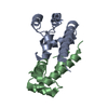 1g8qS S: Starting model for refinement C: citing same article ( |
|---|---|
| Similar structure data |

-Links
 PDBj
PDBj- Assembly
| Deposited unit | 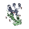
| ||||||||
|---|---|---|---|---|---|---|---|---|---|
| 1 |
| ||||||||
| Unit cell |
|
-Components
| #1: Protein | Mass: 11182.417 Da / Num. of mol.: 2 Source method: isolated from a genetically manipulated source Source: (gene. exp.) Homo sapiens (human) / Gene: CD81, TAPA1, TSPAN28 / Plasmid: pHLSec / Cell line (production host): HEK293T / Production host: Homo sapiens (human) / References: UniProt: P60033#2: Chemical | ChemComp-EDO /   Mass: 62.068 Da / Num. of mol.: 7 / Source method: obtained synthetically / Formula: C2H6O2 Mass: 62.068 Da / Num. of mol.: 7 / Source method: obtained synthetically / Formula: C2H6O2#3: Water | ChemComp-HOH / |  Mass: 18.015 Da / Num. of mol.: 194 / Source method: isolated from a natural source / Formula: H2O Mass: 18.015 Da / Num. of mol.: 194 / Source method: isolated from a natural source / Formula: H2OHas protein modification | Y | |
|---|
-Experimental details
-Experiment
| Experiment | Method: X-RAY DIFFRACTION / Number of used crystals: 1 |
|---|
- Sample preparation
| Crystal | Density Matthews: 1.87 Å3/Da / Density % sol: 34.4 % / Description: excellent diffractor - rodlike shape |
|---|---|
| Crystal grow | Temperature: 294.15 K / Method: vapor diffusion, sitting drop / pH: 5 Details: Protein: 10 mg/ml Buffer: 0.1 M MIB pH 5.0, 25% w/v PEG 1500 grown in presence of synthetic claudin-I long-extracellular-loop (CLDN1-EL1) - but not visible in electron density. cryo- ...Details: Protein: 10 mg/ml Buffer: 0.1 M MIB pH 5.0, 25% w/v PEG 1500 grown in presence of synthetic claudin-I long-extracellular-loop (CLDN1-EL1) - but not visible in electron density. cryo-protectant: 25% glycerol + peptide |
-Data collection
| Diffraction | Mean temperature: 100 K |
|---|---|
| Diffraction source | Source: SYNCHROTRON / Site: ESRF  / Beamline: ID23-1 / Wavelength: 0.972499 Å / Beamline: ID23-1 / Wavelength: 0.972499 Å |
| Detector | Type: DECTRIS PILATUS 6M / Detector: PIXEL / Date: Nov 23, 2012 |
| Radiation | Protocol: SINGLE WAVELENGTH / Monochromatic (M) / Laue (L): M / Scattering type: x-ray |
| Radiation wavelength | Wavelength: 0.972499 Å / Relative weight: 1 |
| Reflection | Resolution: 1.28→26 Å / Num. obs: 38224 / % possible obs: 88.6 % / Redundancy: 5.2 % / Biso Wilson estimate: 16.1 Å2 / Rmerge(I) obs: 0.062 / Net I/σ(I): 21.6 |
| Reflection shell | Resolution: 1.28→1.3 Å / Redundancy: 3.8 % / Rmerge(I) obs: 0.38 / Mean I/σ(I) obs: 3.4 / CC1/2: 0.934 / % possible all: 40.9 |
- Processing
| Software |
| |||||||||||||||||||||||||||||||||||||||||||||||||||||||||||||||||||||||||||||||||||||||||||||||||||||||||
|---|---|---|---|---|---|---|---|---|---|---|---|---|---|---|---|---|---|---|---|---|---|---|---|---|---|---|---|---|---|---|---|---|---|---|---|---|---|---|---|---|---|---|---|---|---|---|---|---|---|---|---|---|---|---|---|---|---|---|---|---|---|---|---|---|---|---|---|---|---|---|---|---|---|---|---|---|---|---|---|---|---|---|---|---|---|---|---|---|---|---|---|---|---|---|---|---|---|---|---|---|---|---|---|---|---|---|
| Refinement | Method to determine structure: MOLECULAR REPLACEMENT Starting model: 1G8Q Resolution: 1.28→25.997 Å / SU ML: 0.11 / Cross valid method: FREE R-VALUE / σ(F): 1.4 / Phase error: 20.02
| |||||||||||||||||||||||||||||||||||||||||||||||||||||||||||||||||||||||||||||||||||||||||||||||||||||||||
| Solvent computation | Shrinkage radii: 0.9 Å / VDW probe radii: 1.11 Å | |||||||||||||||||||||||||||||||||||||||||||||||||||||||||||||||||||||||||||||||||||||||||||||||||||||||||
| Refinement step | Cycle: LAST / Resolution: 1.28→25.997 Å
| |||||||||||||||||||||||||||||||||||||||||||||||||||||||||||||||||||||||||||||||||||||||||||||||||||||||||
| Refine LS restraints |
| |||||||||||||||||||||||||||||||||||||||||||||||||||||||||||||||||||||||||||||||||||||||||||||||||||||||||
| LS refinement shell |
|
