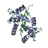
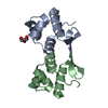
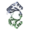
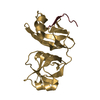
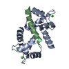
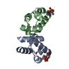
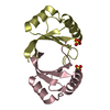
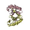
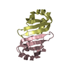
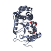
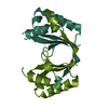
Entry Database : PDB / ID : 3dvmTitle Crystal Structure of Ca2+/CaM-CaV2.1 IQ domain complex Calmodulin Voltage-dependent P/Q-type calcium channel subunit alpha-1A Keywords / / / / / / / Function / homology Function Domain/homology Component
/ / / / / / / / / / / / / / / / / / / / / / / / / / / / / / / / / / / / / / / / / / / / / / / / / / / / / / / / / / / / / / / / / / / / / / / / / / / / / / / / / / / / / / / / / / / / / / / / / / / / / / / / / / / / / / / / / / / / / / / / / / / / / / / / / / / / / / / / / / / Biological species Homo sapiens (human)Oryctolagus cuniculus (rabbit)Method / / / Resolution : 2.6 Å Authors Kim, E.Y. / Rumpf, C.H. / Fujiwara, Y. / Cooley, E.S. / Van Petegem, F. / Minor, D.L. Journal : Structure / Year : 2008Title : Structures of Ca(V)2 Ca(2+)/CaM-IQ Domain Complexes Reveal Binding Modes that Underlie Calcium-Dependent Inactivation and Facilitation.Authors : Kim, E.Y. / Rumpf, C.H. / Fujiwara, Y. / Cooley, E.S. / Van Petegem, F. / Minor, D.L. History Deposition Jul 18, 2008 Deposition site / Processing site Revision 1.0 Nov 4, 2008 Provider / Type Revision 1.1 Jul 13, 2011 Group / Version format complianceRevision 1.2 Oct 25, 2017 Group / Category Revision 1.3 Feb 21, 2024 Group / Database references / Derived calculationsCategory chem_comp_atom / chem_comp_bond ... chem_comp_atom / chem_comp_bond / database_2 / struct_ref_seq_dif / struct_site Item _database_2.pdbx_DOI / _database_2.pdbx_database_accession ... _database_2.pdbx_DOI / _database_2.pdbx_database_accession / _struct_ref_seq_dif.details / _struct_site.pdbx_auth_asym_id / _struct_site.pdbx_auth_comp_id / _struct_site.pdbx_auth_seq_id
Show all Show less
 Movie
Movie Controller


 Open data
 Basic information
 Components
 Keywords
 Function and homology information
 Homo sapiens (human)
 X-RAY DIFFRACTION /
  Authors
 Citation
 Structure visualization
 Downloads & links
 Other downloads
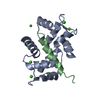

 PDBj
PDBj


 Assembly
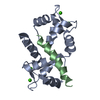


 Mass: 40.078 Da / Num. of mol.: 4 / Source method: obtained synthetically / Formula: Ca
 Mass: 18.015 Da / Num. of mol.: 10 / Source method: isolated from a natural source / Formula: H2O
 Sample preparation
 Processing