 Movie
Movie Controller

[English] 日本語
 Yorodumi
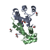
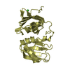
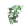
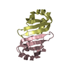
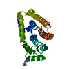
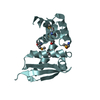
- PDB-1fia: CRYSTAL STRUCTURE OF THE FACTOR FOR INVERSION STIMULATION FIS AT ... -
+ Open data

- Basic information
| Entry | Database: PDB / ID: 1fia | ||||||
|---|---|---|---|---|---|---|---|
| Title | CRYSTAL STRUCTURE OF THE FACTOR FOR INVERSION STIMULATION FIS AT 2.0 ANGSTROMS RESOLUTION | ||||||
 Components Components | FACTOR FOR INVERSION STIMULATION (FIS) | ||||||
 Keywords Keywords | DNA BINDING PROTEIN / DNA-BINDING PROTEIN | ||||||
| Function / homology |  Function and homology information Function and homology informationinvertasome / positive regulation of DNA recombination / sequence-specific DNA binding, bending / provirus excision / nucleoid / DNA-binding transcription repressor activity / DNA-binding transcription activator activity / chromosome organization / core promoter sequence-specific DNA binding / protein-DNA complex ...invertasome / positive regulation of DNA recombination / sequence-specific DNA binding, bending / provirus excision / nucleoid / DNA-binding transcription repressor activity / DNA-binding transcription activator activity / chromosome organization / core promoter sequence-specific DNA binding / protein-DNA complex / response to radiation / nucleosome / sequence-specific DNA binding / transcription cis-regulatory region binding / regulation of DNA-templated transcription / DNA-templated transcription / protein homodimerization activity / DNA binding / cytosol Similarity search - Function | ||||||
| Biological species |  | ||||||
| Method |  X-RAY DIFFRACTION / Resolution: 2 Å X-RAY DIFFRACTION / Resolution: 2 Å | ||||||
 Authors Authors | Kostrewa, D. / Granzin, J. / Choe, H.-W. / Labahn, J. / Saenger, W. | ||||||
 Citation Citation | Journal: J.Mol.Biol. / Year: 1992 Title: Crystal structure of the factor for inversion stimulation FIS at 2.0 A resolution. Authors: Kostrewa, D. / Granzin, J. / Stock, D. / Choe, H.W. / Labahn, J. / Saenger, W. #1: Journal: Nature / Year: 1991Title: Three-Dimensional Structure of the E. Coli DNA-Binding Protein FIS Authors: Kostrewa, D. / Granzin, J. / Koch, C. / Choe, H.-W. / Raghunathan, S. / Wolf, W. / Labahn, J. / Kahmann, R. / Saenger, W. #2: Journal: J.Mol.Biol. / Year: 1989Title: Crystallization of the DNA-Binding Escherichia Coli Protein FIS Authors: Choe, H.-W. / Labahn, J. / Itoh, S. / Koch, C. / Kahmann, R. / Saenger, W. | ||||||
| History |
|
- Structure visualization
| Structure viewer | Molecule: MolmilJmol/JSmol |
|---|
- Downloads & links
-Download
| PDBx/mmCIF format | 1fia.cif.gz | 43.3 KB | Display | PDBx/mmCIF format |
|---|---|---|---|---|
| PDB format | pdb1fia.ent.gz | 31.6 KB | Display | PDB format |
| PDBx/mmJSON format | 1fia.json.gz | Tree view | PDBx/mmJSON format | |
| Others |  Other downloads Other downloads |
-Validation report
| Arichive directory | https://data.pdbj.org/pub/pdb/validation_reports/fi/1fiaftp://data.pdbj.org/pub/pdb/validation_reports/fi/1fia | HTTPS FTP |
|---|
-Related structure data
| Similar structure data |
|---|


-Links
 PDBj
PDBj- Assembly
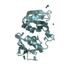
| Deposited unit | 
| ||||||||
|---|---|---|---|---|---|---|---|---|---|
| 1 |
| ||||||||
| Unit cell |
| ||||||||
| Atom site foot note | 1: RESIDUES VAL 10 AND LYS 25 IN BOTH CHAINS WERE MODELLED AS ALANINE. |
-Components
| #1: Protein | Mass: 11252.918 Da / Num. of mol.: 2 Source method: isolated from a genetically manipulated source Source: (gene. exp.) #2: Water | ChemComp-HOH / |  Mass: 18.015 Da / Num. of mol.: 82 / Source method: isolated from a natural source / Formula: H2O Mass: 18.015 Da / Num. of mol.: 82 / Source method: isolated from a natural source / Formula: H2OCompound details | HELICES C AND D OF EACH CHAIN FORM THE HELIX-TURN-HELIX MOTIF FOR DNA BINDING. | |
|---|
-Experimental details
-Experiment
| Experiment | Method: X-RAY DIFFRACTION |
|---|
- Sample preparation
| Crystal | Density Matthews: 2.14 Å3/Da / Density % sol: 42.46 % | ||||||||||||||||||||||||||||||||||||
|---|---|---|---|---|---|---|---|---|---|---|---|---|---|---|---|---|---|---|---|---|---|---|---|---|---|---|---|---|---|---|---|---|---|---|---|---|---|
| Crystal grow | *PLUS pH: 8.2 / Method: vapor diffusion, sitting drop | ||||||||||||||||||||||||||||||||||||
| Components of the solutions | *PLUS
|
-Data collection
| Radiation | Scattering type: x-ray |
|---|---|
| Radiation wavelength | Relative weight: 1 |
| Reflection | *PLUS Highest resolution: 2 Å / Num. all: 12719 / Num. obs: 8221 / % possible obs: 92 % / Num. measured all: 44050 |
- Processing
| Software | Name: TNT / Classification: refinement | ||||||||||||||||||||||||||||||
|---|---|---|---|---|---|---|---|---|---|---|---|---|---|---|---|---|---|---|---|---|---|---|---|---|---|---|---|---|---|---|---|
| Refinement | Rfactor obs: 0.192 / Highest resolution: 2 Å | ||||||||||||||||||||||||||||||
| Refinement step | Cycle: LAST / Highest resolution: 2 Å
| ||||||||||||||||||||||||||||||
| Refine LS restraints |
| ||||||||||||||||||||||||||||||
| Refinement | *PLUS Num. reflection all: 12719 | ||||||||||||||||||||||||||||||
| Solvent computation | *PLUS | ||||||||||||||||||||||||||||||
| Displacement parameters | *PLUS | ||||||||||||||||||||||||||||||
| Refine LS restraints | *PLUS
|
