 Movie
Movie Controller

[English] 日本語
 Yorodumi
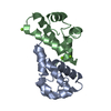
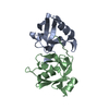
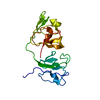
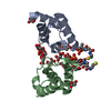
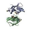
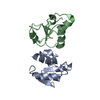
- PDB-1g8q: CRYSTAL STRUCTURE OF HUMAN CD81 EXTRACELLULAR DOMAIN, A RECEPTOR ... -
+ Open data

- Basic information
| Entry | Database: PDB / ID: 1g8q | ||||||
|---|---|---|---|---|---|---|---|
| Title | CRYSTAL STRUCTURE OF HUMAN CD81 EXTRACELLULAR DOMAIN, A RECEPTOR FOR HEPATITIS C VIRUS | ||||||
 Components Components | CD81 ANTIGEN, EXTRACELLULAR DOMAIN | ||||||
 Keywords Keywords | IMMUNE SYSTEM / Alpha Helical | ||||||
| Function / homology |  Function and homology information Function and homology informationpositive regulation of adaptive immune memory response / positive regulation of protein catabolic process in the vacuole / CD4-positive, alpha-beta T cell costimulation / osteoclast fusion / positive regulation of B cell receptor signaling pathway / myoblast fusion involved in skeletal muscle regeneration / positive regulation of T cell activation via T cell receptor contact with antigen bound to MHC molecule on antigen presenting cell / positive regulation of inflammatory response to antigenic stimulus / regulation of macrophage migration / macrophage fusion ...positive regulation of adaptive immune memory response / positive regulation of protein catabolic process in the vacuole / CD4-positive, alpha-beta T cell costimulation / osteoclast fusion / positive regulation of B cell receptor signaling pathway / myoblast fusion involved in skeletal muscle regeneration / positive regulation of T cell activation via T cell receptor contact with antigen bound to MHC molecule on antigen presenting cell / positive regulation of inflammatory response to antigenic stimulus / regulation of macrophage migration / macrophage fusion / humoral immune response mediated by circulating immunoglobulin / immunological synapse formation / tetraspanin-enriched microdomain / positive regulation of T-helper 2 cell cytokine production / transferrin receptor binding / protein localization to lysosome / MHC class II protein binding / positive regulation of protein exit from endoplasmic reticulum / positive regulation of CD4-positive, alpha-beta T cell proliferation / positive regulation of T cell receptor signaling pathway / cholesterol binding / immunological synapse / cellular response to low-density lipoprotein particle stimulus / positive regulation of B cell proliferation / positive regulation of receptor clustering / Regulation of Complement cascade / basal plasma membrane / protein localization to plasma membrane / regulation of protein stability / receptor internalization / integrin binding / Immunoregulatory interactions between a Lymphoid and a non-Lymphoid cell / MHC class II protein complex binding / virus receptor activity / vesicle / basolateral plasma membrane / positive regulation of MAPK cascade / focal adhesion / positive regulation of transcription by RNA polymerase II / extracellular exosome / membrane / plasma membrane Similarity search - Function | ||||||
| Biological species |  Homo sapiens (human) Homo sapiens (human) | ||||||
| Method |  X-RAY DIFFRACTION / SYNCHROTRON / MIR / Resolution: 1.6 Å X-RAY DIFFRACTION / SYNCHROTRON / MIR / Resolution: 1.6 Å | ||||||
 Authors Authors | Kitadokoro, K. / Bolognesi, M. / Bordo, D. / Grandi, G. / Galli, G. / Petracca, R. / Falugi, F. | ||||||
 Citation Citation | Journal: EMBO J. / Year: 2001 Title: CD81 extracellular domain 3D structure: insight into the tetraspanin superfamily structural motifs. Authors: Kitadokoro, K. / Bordo, D. / Galli, G. / Petracca, R. / Falugi, F. / Abrignani, S. / Grandi, G. / Bolognesi, M. | ||||||
| History |
|
- Structure visualization
| Structure viewer | Molecule: MolmilJmol/JSmol |
|---|
- Downloads & links
-Download
| PDBx/mmCIF format | 1g8q.cif.gz | 50.7 KB | Display | PDBx/mmCIF format |
|---|---|---|---|---|
| PDB format | pdb1g8q.ent.gz | 36.7 KB | Display | PDB format |
| PDBx/mmJSON format | 1g8q.json.gz | Tree view | PDBx/mmJSON format | |
| Others |  Other downloads Other downloads |
-Validation report
| Arichive directory | https://data.pdbj.org/pub/pdb/validation_reports/g8/1g8qftp://data.pdbj.org/pub/pdb/validation_reports/g8/1g8q | HTTPS FTP |
|---|
-Related structure data
| Similar structure data |
|---|


-Links
 PDBj
PDBj- Assembly
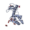
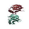
| Deposited unit | 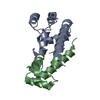
| ||||||||||
|---|---|---|---|---|---|---|---|---|---|---|---|
| 1 |
| ||||||||||
| Unit cell |
|
-Components
| #1: Protein | Mass: 9917.077 Da / Num. of mol.: 2 / Fragment: EXTRACELLULAR DOMAIN Source method: isolated from a genetically manipulated source Source: (gene. exp.) Homo sapiens (human) / Plasmid: PEZZ18 / Production host:  #2: Water | ChemComp-HOH / |  Mass: 18.015 Da / Num. of mol.: 194 / Source method: isolated from a natural source / Formula: H2O Mass: 18.015 Da / Num. of mol.: 194 / Source method: isolated from a natural source / Formula: H2OHas protein modification | Y | |
|---|
-Experimental details
-Experiment
| Experiment | Method: X-RAY DIFFRACTION / Number of used crystals: 1 |
|---|
- Sample preparation
| Crystal | Density Matthews: 2.25 Å3/Da / Density % sol: 45.28 % | ||||||||||||||||||||||||||||||
|---|---|---|---|---|---|---|---|---|---|---|---|---|---|---|---|---|---|---|---|---|---|---|---|---|---|---|---|---|---|---|---|
| Crystal grow | Temperature: 298 K / Method: vapor diffusion, sitting drop / pH: 6 Details: PEG 4000, MES, NaCl, pH 6.0, VAPOR DIFFUSION, SITTING DROP, temperature 298K | ||||||||||||||||||||||||||||||
| Crystal grow | *PLUS Temperature: 293 K | ||||||||||||||||||||||||||||||
| Components of the solutions | *PLUS
|
-Data collection
| Diffraction | Mean temperature: 100 K |
|---|---|
| Diffraction source | Source: SYNCHROTRON / Site: ESRF  / Type: ESRF / Wavelength: 0.93 Å / Type: ESRF / Wavelength: 0.93 Å |
| Detector | Type: MARRESEARCH / Detector: CCD / Date: Nov 19, 1999 |
| Radiation | Monochromator: Sagitally focusing Ge(220) and a multilayer / Protocol: SINGLE WAVELENGTH / Monochromatic (M) / Laue (L): M / Scattering type: x-ray |
| Radiation wavelength | Wavelength: 0.93 Å / Relative weight: 1 |
| Reflection | Resolution: 1.6→50 Å / Num. all: 36910 / Num. obs: 21557 / % possible obs: 98 % / Observed criterion σ(F): 2 / Observed criterion σ(I): 1 / Redundancy: 6.9 % / Biso Wilson estimate: 26.3 Å2 / Rmerge(I) obs: 0.038 / Net I/σ(I): 5.2 |
| Reflection shell | Resolution: 1.6→1.66 Å / Redundancy: 2 % / Rmerge(I) obs: 0.303 / Num. unique all: 2249 / Rsym value: 0.314 / % possible all: 98 |
| Reflection shell | *PLUS % possible obs: 93.1 % |
- Processing
| Software |
| |||||||||||||||||||||||||
|---|---|---|---|---|---|---|---|---|---|---|---|---|---|---|---|---|---|---|---|---|---|---|---|---|---|---|
| Refinement | Method to determine structure: MIR / Resolution: 1.6→20 Å / Isotropic thermal model: Isotropic / Cross valid method: THROUGHOUT / σ(F): 0 / σ(I): 0 / Stereochemistry target values: Engh & Huber
| |||||||||||||||||||||||||
| Refinement step | Cycle: LAST / Resolution: 1.6→20 Å
| |||||||||||||||||||||||||
| Refine LS restraints | Type: refmac / Dev ideal: 0.022 | |||||||||||||||||||||||||
| LS refinement shell | Resolution: 1.6→1.673 Å / Rfactor Rfree error: 0.082
| |||||||||||||||||||||||||
| Software | *PLUS Name: REFMAC / Classification: refinement | |||||||||||||||||||||||||
| Refinement | *PLUS Lowest resolution: 20 Å / σ(F): 0 / % reflection Rfree: 4.8 % / Rfactor obs: 0.187 | |||||||||||||||||||||||||
| Solvent computation | *PLUS | |||||||||||||||||||||||||
| Displacement parameters | *PLUS Biso mean: 35.2 Å2 | |||||||||||||||||||||||||
| Refine LS restraints | *PLUS
|
