 Movie
Movie Controller

[English] 日本語
 Yorodumi
- PDB-4k6x: Crystal structure of disulfide oxidoreductase from Mycobacterium ... -
+ Open data

- Basic information
| Entry | Database: PDB / ID: 4k6x | ||||||
|---|---|---|---|---|---|---|---|
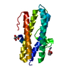
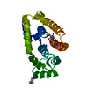
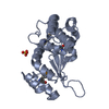
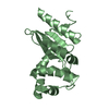
| Title | Crystal structure of disulfide oxidoreductase from Mycobacterium tuberculosis | ||||||
 Components Components | Disulfide oxidoreductase | ||||||
 Keywords Keywords | OXIDOREDUCTASE / DsbA / Disulfide oxidase / VKOR / Thioredoxin fold | ||||||
| Function / homology |  Function and homology information Function and homology informationpeptidoglycan-based cell wall / oxidoreductase activity / cell surface / plasma membrane Similarity search - Function | ||||||
| Biological species |   Mycobacterium tuberculosis (bacteria) Mycobacterium tuberculosis (bacteria) | ||||||
| Method |  X-RAY DIFFRACTION / SYNCHROTRON / MOLECULAR REPLACEMENT / Resolution: 1.972 Å X-RAY DIFFRACTION / SYNCHROTRON / MOLECULAR REPLACEMENT / Resolution: 1.972 Å | ||||||
 Authors Authors | Premkumar, L. / Martin, J.L. | ||||||
 Citation Citation | Journal: Acta Crystallogr.,Sect.D / Year: 2013 Title: Rv2969c, essential for optimal growth in Mycobacterium tuberculosis, is a DsbA-like enzyme that interacts with VKOR-derived peptides and has atypical features of DsbA-like disulfide oxidases. Authors: Premkumar, L. / Heras, B. / Duprez, W. / Walden, P. / Halili, M. / Kurth, F. / Fairlie, D.P. / Martin, J.L. | ||||||
| History |
|
- Structure visualization
| Structure viewer | Molecule: MolmilJmol/JSmol |
|---|
- Downloads & links
-Download
| PDBx/mmCIF format | 4k6x.cif.gz | 233.5 KB | Display | PDBx/mmCIF format |
|---|---|---|---|---|
| PDB format | pdb4k6x.ent.gz | 192.3 KB | Display | PDB format |
| PDBx/mmJSON format | 4k6x.json.gz | Tree view | PDBx/mmJSON format | |
| Others |  Other downloads Other downloads |
-Validation report
| Arichive directory | https://data.pdbj.org/pub/pdb/validation_reports/k6/4k6xftp://data.pdbj.org/pub/pdb/validation_reports/k6/4k6x | HTTPS FTP |
|---|
-Related structure data
| Similar structure data |
|---|


-Links
 PDBj
PDBj- Assembly
| Deposited unit | 
| |||||||||||||||
|---|---|---|---|---|---|---|---|---|---|---|---|---|---|---|---|---|
| 1 | 
| |||||||||||||||
| 2 | 
| |||||||||||||||
| Unit cell |
| |||||||||||||||
| Components on special symmetry positions |
|
-Components
| #1: Protein | Mass: 22275.945 Da / Num. of mol.: 2 / Fragment: UNP residues 52-261 Source method: isolated from a genetically manipulated source Source: (gene. exp.) Mycobacterium tuberculosis (bacteria) / Gene: Rv2969c / Production host: #2: Chemical | ChemComp-EPE / |   Mass: 238.305 Da / Num. of mol.: 1 / Source method: obtained synthetically / Formula: C8H18N2O4S / Comment: pH buffer*YM Mass: 238.305 Da / Num. of mol.: 1 / Source method: obtained synthetically / Formula: C8H18N2O4S / Comment: pH buffer*YM#3: Chemical | ChemComp-DIO / |   Mass: 88.105 Da / Num. of mol.: 1 / Source method: obtained synthetically / Formula: C4H8O2 Mass: 88.105 Da / Num. of mol.: 1 / Source method: obtained synthetically / Formula: C4H8O2#4: Water | ChemComp-HOH / |  Mass: 18.015 Da / Num. of mol.: 412 / Source method: isolated from a natural source / Formula: H2O Mass: 18.015 Da / Num. of mol.: 412 / Source method: isolated from a natural source / Formula: H2OHas protein modification | Y | |
|---|
-Experimental details
-Experiment
| Experiment | Method: X-RAY DIFFRACTION / Number of used crystals: 1 |
|---|
- Sample preparation
| Crystal | Density Matthews: 2.77 Å3/Da / Density % sol: 50.61 % |
|---|---|
| Crystal grow | Temperature: 293 K / Method: vapor diffusion, hanging drop / pH: 5.5 Details: Na Malonate, 1,4-dioxane and polyvinylpyrrolidone, pH 5.5, VAPOR DIFFUSION, HANGING DROP, temperature 293K |
-Data collection
| Diffraction source | Source: SYNCHROTRON / Site: Australian Synchrotron  / Beamline: MX2 / Wavelength: 0.9537 Å / Beamline: MX2 / Wavelength: 0.9537 Å | ||||||||||||||||||
|---|---|---|---|---|---|---|---|---|---|---|---|---|---|---|---|---|---|---|---|
| Detector | Type: ADSC QUANTUM 315r / Detector: CCD / Date: Apr 2, 2012 | ||||||||||||||||||
| Radiation | Monochromator: si(111) / Protocol: SINGLE WAVELENGTH / Monochromatic (M) / Laue (L): M / Scattering type: x-ray | ||||||||||||||||||
| Radiation wavelength | Wavelength: 0.9537 Å / Relative weight: 1 | ||||||||||||||||||
| Reflection | Resolution: 1.97→85.6 Å / Num. all: 35185 / Num. obs: 35185 / % possible obs: 98.9 % / Observed criterion σ(F): 2 / Observed criterion σ(I): 2 / Redundancy: 8.1 % / Biso Wilson estimate: 23.6 Å2 / Rmerge(I) obs: 0.096 / Net I/σ(I): 14.9 | ||||||||||||||||||
| Reflection shell |
|
- Processing
| Software |
| ||||||||||||||||||||||||||||||||||||||||||||||||||||||||||||||||||||||||||||||||||||||||||||||||||
|---|---|---|---|---|---|---|---|---|---|---|---|---|---|---|---|---|---|---|---|---|---|---|---|---|---|---|---|---|---|---|---|---|---|---|---|---|---|---|---|---|---|---|---|---|---|---|---|---|---|---|---|---|---|---|---|---|---|---|---|---|---|---|---|---|---|---|---|---|---|---|---|---|---|---|---|---|---|---|---|---|---|---|---|---|---|---|---|---|---|---|---|---|---|---|---|---|---|---|---|
| Refinement | Method to determine structure: MOLECULAR REPLACEMENT / Resolution: 1.972→59.324 Å / SU ML: 0.19 / σ(F): 1.37 / Phase error: 17.65 / Stereochemistry target values: ML
| ||||||||||||||||||||||||||||||||||||||||||||||||||||||||||||||||||||||||||||||||||||||||||||||||||
| Solvent computation | Shrinkage radii: 0.7 Å / VDW probe radii: 1.1 Å / Solvent model: FLAT BULK SOLVENT MODEL | ||||||||||||||||||||||||||||||||||||||||||||||||||||||||||||||||||||||||||||||||||||||||||||||||||
| Refinement step | Cycle: LAST / Resolution: 1.972→59.324 Å
| ||||||||||||||||||||||||||||||||||||||||||||||||||||||||||||||||||||||||||||||||||||||||||||||||||
| Refine LS restraints |
| ||||||||||||||||||||||||||||||||||||||||||||||||||||||||||||||||||||||||||||||||||||||||||||||||||
| LS refinement shell |
| ||||||||||||||||||||||||||||||||||||||||||||||||||||||||||||||||||||||||||||||||||||||||||||||||||
| Refinement TLS params. | Method: refined / Refine-ID: X-RAY DIFFRACTION
| ||||||||||||||||||||||||||||||||||||||||||||||||||||||||||||||||||||||||||||||||||||||||||||||||||
| Refinement TLS group |
|
