[English] 日本語
Yorodumi
- PDB-2rem: Crystal Structure of oxidoreductase DsbA from Xylella fastidiosa -

+
Open data


ID or keywords:

Loading...

-
Basic information

Entry
Database: PDB / ID: 2rem
TitleCrystal Structure of oxidoreductase DsbA from Xylella fastidiosa
Components
  • 8 residue peptide
  • Disulfide oxidoreductase
KeywordsOXIDOREDUCTASE / disulfide oxidoreductase / DSBA / thioredoxin fold / redox-active center
Function / homology
Function and homology information


cell redox homeostasis / periplasmic space
Similarity search - Function
Thioredoxin / Thioredoxin-like fold / Thiol:disulphide interchange protein DsbA/DsbL / Thioredoxin, conserved site / Thioredoxin family active site. / Thioredoxin domain profile. / Thioredoxin domain / Glutaredoxin / Glutaredoxin / Thioredoxin-like superfamily ...Thioredoxin / Thioredoxin-like fold / Thiol:disulphide interchange protein DsbA/DsbL / Thioredoxin, conserved site / Thioredoxin family active site. / Thioredoxin domain profile. / Thioredoxin domain / Glutaredoxin / Glutaredoxin / Thioredoxin-like superfamily / 3-Layer(aba) Sandwich / Alpha Beta
Similarity search - Domain/homology
Thiol:disulfide interchange protein
Similarity search - Component
Biological speciesXylella fastidiosa (bacteria)
MethodX-RAY DIFFRACTION / SYNCHROTRON / SAD / Resolution: 1.9 Å
AuthorsRinaldi, F.C. / Guimaraes, B.G.
CitationJournal: To be Published
Title: Residues substitution in the active site of DSBA may compensate for the lack of the canonical motif CPHC
Authors: Rinaldi, F.C. / Guimaraes, B.G.
History
DepositionSep 26, 2007Deposition site: RCSB / Processing site: RCSB
Revision 1.0Oct 7, 2008Provider: repository / Type: Initial release
Revision 1.1Jul 13, 2011Group: Version format compliance
Revision 1.2Oct 25, 2017Group: Advisory / Refinement description / Category: pdbx_unobs_or_zero_occ_atoms / software
Revision 1.3Feb 21, 2024Group: Advisory / Data collection / Database references
Category: chem_comp_atom / chem_comp_bond ...chem_comp_atom / chem_comp_bond / database_2 / pdbx_unobs_or_zero_occ_atoms
Item: _database_2.pdbx_DOI / _database_2.pdbx_database_accession

-
Structure visualization

Structure viewerMolecule:
MolmilJmol/JSmol

Downloads & links

-
Assembly

Deposited unit
A: Disulfide oxidoreductase
B: Disulfide oxidoreductase
C: Disulfide oxidoreductase
T: 8 residue peptide


Theoretical massNumber of molelcules
Total (without water)64,6994
Polymers64,6994
Non-polymers00
Water8,737485
1
A: Disulfide oxidoreductase


Theoretical massNumber of molelcules
Total (without water)21,3331
Polymers21,3331
Non-polymers00
Water181
TypeNameSymmetry operationNumber
identity operation1_555x,y,z1
MethodPISA
2
B: Disulfide oxidoreductase


Theoretical massNumber of molelcules
Total (without water)21,3331
Polymers21,3331
Non-polymers00
Water181
TypeNameSymmetry operationNumber
identity operation1_555x,y,z1
MethodPISA
3
C: Disulfide oxidoreductase
T: 8 residue peptide


Theoretical massNumber of molelcules
Total (without water)22,0322
Polymers22,0322
Non-polymers00
Water362
TypeNameSymmetry operationNumber
identity operation1_555x,y,z1
MethodPISA
Unit cell
Length a, b, c (Å)200.117, 41.722, 79.807
Angle α, β, γ (deg.)90.000, 95.870, 90.000
Int Tables number5
Space group name H-MC121

-
Components

#1: Protein Disulfide oxidoreductase


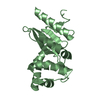
Mass: 21333.254 Da / Num. of mol.: 3
Source method: isolated from a genetically manipulated source
Source: (gene. exp.) Xylella fastidiosa (bacteria) / Gene: XF1436 / Plasmid: pET28a(+) / Production host: Escherichia coli (E. coli) / Strain (production host): C43(DE3)
References: UniProt: Q9PDE3, protein-disulfide reductase (glutathione)
#2: Protein/peptide 8 residue peptide


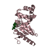
Mass: 698.854 Da / Num. of mol.: 1
Source method: isolated from a genetically manipulated source
Source: (gene. exp.) Xylella fastidiosa (bacteria)
Description: CHAIN T IS DERIVED FROM THE EXPRESSION SYSTEM THAT WAS CO-PURIFIED WITH THE PROTEIN (XFDSBA)
Plasmid: pET28a(+) / Production host: Escherichia coli (E. coli) / Strain (production host): C43(DE3)
#3: Water ChemComp-HOH / water


Mass: 18.015 Da / Num. of mol.: 485 / Source method: isolated from a natural source / Formula: H2O
Sequence detailsTHE ACTUAL SEQUENCE OF CHAIN T IS UNKNOWN. THESE RESIDUES WERE MODELED AS UNK DUE TO WEAK ELECTRON DENSITY.

-
Experimental details

-
Experiment

ExperimentMethod: X-RAY DIFFRACTION / Number of used crystals: 2

-
Sample preparation

CrystalDensity Matthews: 2.56 Å3/Da / Density % sol: 51.98 %
Crystal growTemperature: 293 K / Method: vapor diffusion, sitting drop / pH: 5
Details: 30% PEG 4000, 0.1M acetate chloride, Guanidine Hydrochloride, pH 5.0, VAPOR DIFFUSION, SITTING DROP, temperature 293K

-
Data collection

Diffraction
IDMean temperature (K)Crystal-ID
11001
21001
Diffraction source
SourceSiteBeamlineIDWavelength (Å)
SYNCHROTRONLNLS D03B-MX111.431
SYNCHROTRONNSLS X26C20.9795
Detector
TypeIDDetectorDate
MAR CCD 165 mm1CCDJan 26, 2005
ADSC QUANTUM 42CCDApr 24, 2006
Radiation
IDMonochromatorProtocolMonochromatic (M) / Laue (L)Scattering typeWavelength-ID
1Single Crystal Monochromator Si(111)SINGLE WAVELENGTHMx-ray1
2Single Crystal Monochromator Si(111)SINGLE WAVELENGTHMx-ray1
Radiation wavelength
IDWavelength (Å)Relative weight
11.4311
20.97951
ReflectionRedundancy: 6.6 % / Av σ(I) over netI: 22.6 / Number: 360459 / Rmerge(I) obs: 0.079 / Χ2: 4.32 / D res high: 1.85 Å / D res low: 50 Å / Num. obs: 54206 / % possible obs: 91.5
Diffraction reflection shell
Highest resolution (Å)Lowest resolution (Å)% possible obs (%)IDRmerge(I) obsChi squaredRedundancy
3.995098.610.0628.6376.4
3.163.9981.510.0535.3636.6
2.763.1699.910.0654.8937.2
2.512.7610010.0854.0087.3
2.332.5110010.1073.4657.3
2.192.3351.410.1593.7975
2.082.1910010.1952.8817.1
1.992.0899.910.2282.5187.3
1.921.9999.510.4663.4256.3
1.851.9283.610.3134.6364.9
ReflectionResolution: 1.9→50 Å / Num. all: 52300 / Num. obs: 52143 / % possible obs: 99.7 % / Observed criterion σ(F): 2 / Observed criterion σ(I): 2 / Redundancy: 3.5 % / Biso Wilson estimate: 21.8 Å2 / Rmerge(I) obs: 0.074 / Χ2: 0.754 / Net I/σ(I): 7.3
Reflection shellResolution: 1.9→1.97 Å / Redundancy: 2.9 % / Rmerge(I) obs: 0.479 / Num. unique all: 5052 / Χ2: 1.366 / % possible all: 98.3

-
Phasing

PhasingMethod: SAD

-
Processing

Software
NameVersionClassificationNB
DENZOdata reduction
SCALEPACKdata scaling
SHARPphasing
REFMACrefinement
PDB_EXTRACT3data extraction
CBASSdata collection
HKL-2000data reduction
HKL-2000data scaling
RefinementMethod to determine structure: SAD / Resolution: 1.9→50 Å / Cor.coef. Fo:Fc: 0.949 / Cor.coef. Fo:Fc free: 0.928 / SU B: 5.699 / SU ML: 0.091 / Cross valid method: THROUGHOUT / σ(F): 2 / σ(I): 2 / ESU R: 0.145 / ESU R Free: 0.137 / Stereochemistry target values: MAXIMUM LIKELIHOOD / Details: HYDROGENS HAVE BEEN ADDED IN THE RIDING POSITIONS
RfactorNum. reflection% reflectionSelection details
Rfree0.224 2657 5.1 %RANDOM
Rwork0.183 ---
all0.204 52367 --
obs0.185 52137 99.56 %-
Solvent computationIon probe radii: 0.8 Å / Shrinkage radii: 0.8 Å / VDW probe radii: 1.4 Å / Solvent model: MASK
Displacement parametersBiso mean: 24.959 Å2
Baniso -1Baniso -2Baniso -3
1--0.43 Å20 Å2-0.36 Å2
2--1.09 Å20 Å2
3----0.73 Å2
Refinement stepCycle: LAST / Resolution: 1.9→50 Å
ProteinNucleic acidLigandSolventTotal
Num. atoms4489 0 0 485 4974
Refine LS restraints
Refine-IDTypeDev idealDev ideal targetNumber
X-RAY DIFFRACTIONr_bond_refined_d0.0110.0224614
X-RAY DIFFRACTIONr_angle_refined_deg1.2881.9396288
X-RAY DIFFRACTIONr_dihedral_angle_1_deg5.4755579
X-RAY DIFFRACTIONr_dihedral_angle_2_deg33.2722.431218
X-RAY DIFFRACTIONr_dihedral_angle_3_deg14.15515664
X-RAY DIFFRACTIONr_dihedral_angle_4_deg20.6411539
X-RAY DIFFRACTIONr_chiral_restr0.0910.2673
X-RAY DIFFRACTIONr_gen_planes_refined0.0060.023655
X-RAY DIFFRACTIONr_nbd_refined0.2070.22654
X-RAY DIFFRACTIONr_nbtor_refined0.3170.23284
X-RAY DIFFRACTIONr_xyhbond_nbd_refined0.1560.2573
X-RAY DIFFRACTIONr_symmetry_vdw_refined0.2730.2113
X-RAY DIFFRACTIONr_symmetry_hbond_refined0.20.232
X-RAY DIFFRACTIONr_mcbond_it0.6881.52904
X-RAY DIFFRACTIONr_mcangle_it1.11624616
X-RAY DIFFRACTIONr_scbond_it2.09831896
X-RAY DIFFRACTIONr_scangle_it3.4084.51668
LS refinement shellResolution: 1.9→1.949 Å / Total num. of bins used: 20
RfactorNum. reflection% reflection
Rfree0.327 178 -
Rwork0.245 3488 -
all-3666 -
obs-3666 96.5 %
Refinement TLS params.

Method: refined / Refine-ID: X-RAY DIFFRACTION

IDL112)L122)L132)L222)L232)L332)S11 (Å °)S12 (Å °)S13 (Å °)S21 (Å °)S22 (Å °)S23 (Å °)S31 (Å °)S32 (Å °)S33 (Å °)T112)T122)T132)T222)T232)T332)Origin x (Å)Origin y (Å)Origin z (Å)
10.9787-0.35331.15011.005-0.68633.431-0.0128-0.06310.03240.0268-0.0065-0.0325-0.03540.02590.0193-0.1486-0.01740.0228-0.18510.0039-0.132437.58946.27484.5213
22.2547-0.17460.86331.0743-1.06363.8989-0.02220.21930.1445-0.02560.08450.1070.004-0.0189-0.0623-0.1611-0.01520.0185-0.09630.042-0.121726.7523-7.093331.5441
33.0357-1.11380.04481.62030.41041.45240.05160.11080.1126-0.0892-0.15560.0679-0.0598-0.23860.104-0.133-0.0087-0.0021-0.1059-0.0075-0.154815.6013-8.2248-21.5007
Refinement TLS group
IDRefine-IDRefine TLS-IDAuth asym-IDLabel asym-IDAuth seq-IDLabel seq-ID
1X-RAY DIFFRACTION1AA4 - 1933 - 192
2X-RAY DIFFRACTION2BB4 - 1903 - 189
3X-RAY DIFFRACTION3CC4 - 1933 - 192

+
About Yorodumi

-
News

-
Feb 9, 2022. New format data for meta-information of EMDB entries

New format data for meta-information of EMDB entries

  • Version 3 of the EMDB header file is now the official format.
  • The previous official version 1.9 will be removed from the archive.

Related info.:EMDB header

External links:wwPDB to switch to version 3 of the EMDB data model

-
Aug 12, 2020. Covid-19 info

Covid-19 info

URL: https://pdbjlvh1.pdbj.org/emnavi/covid19.php

New page: Covid-19 featured information page in EM Navigator.

Related info.:Covid-19 info / Mar 5, 2020. Novel coronavirus structure data

+
Mar 5, 2020. Novel coronavirus structure data

Novel coronavirus structure data

Related info.:Yorodumi Speices / Aug 12, 2020. Covid-19 info

External links:COVID-19 featured content - PDBj / Molecule of the Month (242):Coronavirus Proteases

+
Jan 31, 2019. EMDB accession codes are about to change! (news from PDBe EMDB page)

EMDB accession codes are about to change! (news from PDBe EMDB page)

  • The allocation of 4 digits for EMDB accession codes will soon come to an end. Whilst these codes will remain in use, new EMDB accession codes will include an additional digit and will expand incrementally as the available range of codes is exhausted. The current 4-digit format prefixed with “EMD-” (i.e. EMD-XXXX) will advance to a 5-digit format (i.e. EMD-XXXXX), and so on. It is currently estimated that the 4-digit codes will be depleted around Spring 2019, at which point the 5-digit format will come into force.
  • The EM Navigator/Yorodumi systems omit the EMD- prefix.

Related info.:Q: What is EMD? / ID/Accession-code notation in Yorodumi/EM Navigator

External links:EMDB Accession Codes are Changing Soon! / Contact to PDBj

+
Jul 12, 2017. Major update of PDB

Major update of PDB

  • wwPDB released updated PDB data conforming to the new PDBx/mmCIF dictionary.
  • This is a major update changing the version number from 4 to 5, and with Remediation, in which all the entries are updated.
  • In this update, many items about electron microscopy experimental information are reorganized (e.g. em_software).
  • Now, EM Navigator and Yorodumi are based on the updated data.

External links:wwPDB Remediation / Enriched Model Files Conforming to OneDep Data Standards Now Available in the PDB FTP Archive

-
Yorodumi

Thousand views of thousand structures

  • Yorodumi is a browser for structure data from EMDB, PDB, SASBDB, etc.
  • This page is also the successor to EM Navigator detail page, and also detail information page/front-end page for Omokage search.
  • The word "yorodu" (or yorozu) is an old Japanese word meaning "ten thousand". "mi" (miru) is to see.

Related info.:EMDB / PDB / SASBDB / Comparison of 3 databanks / Yorodumi Search / Aug 31, 2016. New EM Navigator & Yorodumi / Yorodumi Papers / Jmol/JSmol / Function and homology information / Changes in new EM Navigator and Yorodumi

Read more