 Movie
Movie Controller

[English] 日本語
 Yorodumi
- PDB-5lab: Crystal structure of the catalytic domain of human MMP12 complexe... -
+ Open data

- Basic information
| Entry | Database: PDB / ID: 5lab | ||||||
|---|---|---|---|---|---|---|---|
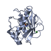
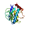
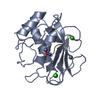
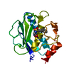
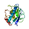
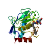
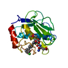
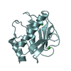
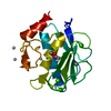
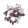
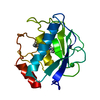
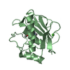
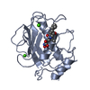
| Title | Crystal structure of the catalytic domain of human MMP12 complexed with the inhibitor NNGH | ||||||
 Components Components | Macrophage metalloelastase | ||||||
 Keywords Keywords | HYDROLASE / HYDROLASE MMP-12 | ||||||
| Function / homology |  Function and homology information Function and homology informationmacrophage elastase / negative regulation of endothelial cell-matrix adhesion / bronchiole development / positive regulation of epithelial cell proliferation involved in wound healing / elastin catabolic process / regulation of defense response to virus by host / positive regulation of type I interferon-mediated signaling pathway / wound healing, spreading of epidermal cells / negative regulation of type I interferon-mediated signaling pathway / lung alveolus development ...macrophage elastase / negative regulation of endothelial cell-matrix adhesion / bronchiole development / positive regulation of epithelial cell proliferation involved in wound healing / elastin catabolic process / regulation of defense response to virus by host / positive regulation of type I interferon-mediated signaling pathway / wound healing, spreading of epidermal cells / negative regulation of type I interferon-mediated signaling pathway / lung alveolus development / response to amyloid-beta / Collagen degradation / collagen catabolic process / positive regulation of interferon-alpha production / extracellular matrix disassembly / core promoter sequence-specific DNA binding / Degradation of the extracellular matrix / collagen binding / extracellular matrix organization / metalloendopeptidase activity / cellular response to virus / protein import into nucleus / extracellular matrix / endopeptidase activity / sequence-specific DNA binding / serine-type endopeptidase activity / calcium ion binding / negative regulation of transcription by RNA polymerase II / positive regulation of transcription by RNA polymerase II / proteolysis / : / extracellular region / zinc ion binding / nucleus / cytoplasm Similarity search - Function | ||||||
| Biological species |  Homo sapiens (human) Homo sapiens (human) | ||||||
| Method |  X-RAY DIFFRACTION / SYNCHROTRON / MOLECULAR REPLACEMENT / Resolution: 1.34 Å X-RAY DIFFRACTION / SYNCHROTRON / MOLECULAR REPLACEMENT / Resolution: 1.34 Å | ||||||
 Authors Authors | Ravera, E. / Calderone, V. / Luchinat, C. | ||||||
 Citation Citation | Journal: To Be Published Title: First-principles calculation of pseudo-contact shifts in a paramagnetic metalloprotein Authors: Benda, L. / Mares, J. / Ravera, E. / Parigi, G. / Luchinat, C. / Kaupp, M. / Vaara, J. #1: Journal: Proc. Natl. Acad. Sci. U.S.A. / Year: 2005Title: Conformational variability of matrix metalloproteinases: beyond a single 3D structure. Authors: Bertini, I. / Calderone, V. / Cosenza, M. / Fragai, M. / Lee, Y.M. / Luchinat, C. / Mangani, S. / Terni, B. / Turano, P. | ||||||
| History |
|
- Structure visualization
| Structure viewer | Molecule: MolmilJmol/JSmol |
|---|
- Downloads & links
-Download
| PDBx/mmCIF format | 5lab.cif.gz | 91.3 KB | Display | PDBx/mmCIF format |
|---|---|---|---|---|
| PDB format | pdb5lab.ent.gz | 67.2 KB | Display | PDB format |
| PDBx/mmJSON format | 5lab.json.gz | Tree view | PDBx/mmJSON format | |
| Others |  Other downloads Other downloads |
-Validation report
| Arichive directory | https://data.pdbj.org/pub/pdb/validation_reports/la/5labftp://data.pdbj.org/pub/pdb/validation_reports/la/5lab | HTTPS FTP |
|---|
-Related structure data
| Related structure data |  1os9S S: Starting model for refinement |
|---|---|
| Similar structure data |


-Links
 PDBj
PDBj


- Assembly
| Deposited unit | 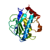
| ||||||||
|---|---|---|---|---|---|---|---|---|---|
| 1 |
| ||||||||
| Unit cell |
| ||||||||
| Components on special symmetry positions |
|
-Components
| #1: Protein | Mass: 17615.670 Da / Num. of mol.: 1 / Fragment: UNP residues 106-263 / Mutation: F171D Source method: isolated from a genetically manipulated source Source: (gene. exp.) Homo sapiens (human) / Gene: MMP12, HME / Production host:  | ||||||||
|---|---|---|---|---|---|---|---|---|---|
| #2: Chemical |   Mass: 65.409 Da / Num. of mol.: 2 / Source method: obtained synthetically / Formula: Zn Mass: 65.409 Da / Num. of mol.: 2 / Source method: obtained synthetically / Formula: Zn#3: Chemical |   Mass: 40.078 Da / Num. of mol.: 3 / Source method: obtained synthetically / Formula: Ca Mass: 40.078 Da / Num. of mol.: 3 / Source method: obtained synthetically / Formula: Ca#4: Chemical | ChemComp-NGH / |   Mass: 316.373 Da / Num. of mol.: 1 / Source method: obtained synthetically / Formula: C13H20N2O5S Mass: 316.373 Da / Num. of mol.: 1 / Source method: obtained synthetically / Formula: C13H20N2O5S#5: Water | ChemComp-HOH / |  Mass: 18.015 Da / Num. of mol.: 236 / Source method: isolated from a natural source / Formula: H2O Mass: 18.015 Da / Num. of mol.: 236 / Source method: isolated from a natural source / Formula: H2OHas protein modification | N | |
-Experimental details
-Experiment
| Experiment | Method: X-RAY DIFFRACTION / Number of used crystals: 1 |
|---|
- Sample preparation
| Crystal | Density Matthews: 2.26 Å3/Da / Density % sol: 45.26 % |
|---|---|
| Crystal grow | Temperature: 293 K / Method: vapor diffusion, sitting drop / pH: 8 Details: Tris, PEG6000, NNGH, pH 8.0, VAPOR DIFFUSION, SITTING DROP, temperature 293K |
-Data collection
| Diffraction | Mean temperature: 100 K |
|---|---|
| Diffraction source | Source: SYNCHROTRON / Site: ELETTRA  / Beamline: 5.2R / Wavelength: 1.2 Å / Beamline: 5.2R / Wavelength: 1.2 Å |
| Detector | Type: MARRESEARCH / Detector: CCD / Date: Oct 5, 2003 |
| Radiation | Monochromator: double-crystal monochromator Si(111) and Si(220) Protocol: SINGLE WAVELENGTH / Monochromatic (M) / Laue (L): M / Scattering type: x-ray |
| Radiation wavelength | Wavelength: 1.2 Å / Relative weight: 1 |
| Reflection | Resolution: 1.34→46.407 Å / Num. obs: 36295 / % possible obs: 98.4 % / Redundancy: 6.2 % / Net I/σ(I): 7.2 |
| Reflection shell | Resolution: 1.34→1.41 Å / Redundancy: 4.8 % / Rmerge(I) obs: 0.308 / Mean I/σ(I) obs: 2.3 / % possible all: 92.6 |
- Processing
| Software |
| ||||||||||||||||||||||||||||||||||||||||||||||||||||||||||||||||||||||||||||||||||||||||||||||||||||||||||||||||||||||||||||||||||||||||||||||||||||||||||||||||||||||||||||||||||||||
|---|---|---|---|---|---|---|---|---|---|---|---|---|---|---|---|---|---|---|---|---|---|---|---|---|---|---|---|---|---|---|---|---|---|---|---|---|---|---|---|---|---|---|---|---|---|---|---|---|---|---|---|---|---|---|---|---|---|---|---|---|---|---|---|---|---|---|---|---|---|---|---|---|---|---|---|---|---|---|---|---|---|---|---|---|---|---|---|---|---|---|---|---|---|---|---|---|---|---|---|---|---|---|---|---|---|---|---|---|---|---|---|---|---|---|---|---|---|---|---|---|---|---|---|---|---|---|---|---|---|---|---|---|---|---|---|---|---|---|---|---|---|---|---|---|---|---|---|---|---|---|---|---|---|---|---|---|---|---|---|---|---|---|---|---|---|---|---|---|---|---|---|---|---|---|---|---|---|---|---|---|---|---|---|
| Refinement | Method to determine structure: MOLECULAR REPLACEMENT Starting model: 1OS9 Resolution: 1.34→20 Å / Cor.coef. Fo:Fc: 0.973 / Cor.coef. Fo:Fc free: 0.963 / SU B: 2.615 / SU ML: 0.046 / Cross valid method: THROUGHOUT / ESU R: 0.059 / ESU R Free: 0.057 / Details: HYDROGENS HAVE BEEN ADDED IN THE RIDING POSITIONS
| ||||||||||||||||||||||||||||||||||||||||||||||||||||||||||||||||||||||||||||||||||||||||||||||||||||||||||||||||||||||||||||||||||||||||||||||||||||||||||||||||||||||||||||||||||||||
| Solvent computation | Ion probe radii: 0.8 Å / Shrinkage radii: 0.8 Å / VDW probe radii: 1.2 Å | ||||||||||||||||||||||||||||||||||||||||||||||||||||||||||||||||||||||||||||||||||||||||||||||||||||||||||||||||||||||||||||||||||||||||||||||||||||||||||||||||||||||||||||||||||||||
| Displacement parameters | Biso mean: 21.754 Å2
| ||||||||||||||||||||||||||||||||||||||||||||||||||||||||||||||||||||||||||||||||||||||||||||||||||||||||||||||||||||||||||||||||||||||||||||||||||||||||||||||||||||||||||||||||||||||
| Refinement step | Cycle: 1 / Resolution: 1.34→20 Å
| ||||||||||||||||||||||||||||||||||||||||||||||||||||||||||||||||||||||||||||||||||||||||||||||||||||||||||||||||||||||||||||||||||||||||||||||||||||||||||||||||||||||||||||||||||||||
| Refine LS restraints |
|