 Movie
Movie Controller

[English] 日本語
 Yorodumi
- PDB-1ros: Crystal structure of MMP-12 complexed to 2-(1,3-dioxo-1,3-dihydro... -
+ Open data

- Basic information
| Entry | Database: PDB / ID: 1ros | ||||||
|---|---|---|---|---|---|---|---|
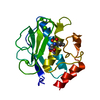
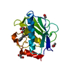
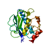
| Title | Crystal structure of MMP-12 complexed to 2-(1,3-dioxo-1,3-dihydro-2H-isoindol-2-yl)ethyl-4-(4-ethoxy[1,1-biphenyl]-4-yl)-4-oxobutanoic acid | ||||||
 Components Components | Macrophage metalloelastase | ||||||
 Keywords Keywords | HYDROLASE / Macrophage metalloelastase / non-zinc chelator / MMP-12 / MMP12 inhibitor | ||||||
| Function / homology |  Function and homology information Function and homology informationmacrophage elastase / negative regulation of endothelial cell-matrix adhesion / bronchiole development / positive regulation of epithelial cell proliferation involved in wound healing / elastin catabolic process / regulation of defense response to virus by host / positive regulation of type I interferon-mediated signaling pathway / wound healing, spreading of epidermal cells / negative regulation of type I interferon-mediated signaling pathway / lung alveolus development ...macrophage elastase / negative regulation of endothelial cell-matrix adhesion / bronchiole development / positive regulation of epithelial cell proliferation involved in wound healing / elastin catabolic process / regulation of defense response to virus by host / positive regulation of type I interferon-mediated signaling pathway / wound healing, spreading of epidermal cells / negative regulation of type I interferon-mediated signaling pathway / lung alveolus development / response to amyloid-beta / Collagen degradation / collagen catabolic process / positive regulation of interferon-alpha production / extracellular matrix disassembly / core promoter sequence-specific DNA binding / Degradation of the extracellular matrix / collagen binding / extracellular matrix organization / metalloendopeptidase activity / cellular response to virus / protein import into nucleus / extracellular matrix / endopeptidase activity / sequence-specific DNA binding / serine-type endopeptidase activity / calcium ion binding / negative regulation of transcription by RNA polymerase II / positive regulation of transcription by RNA polymerase II / proteolysis / : / extracellular region / zinc ion binding / nucleus / cytoplasm Similarity search - Function | ||||||
| Biological species |  Homo sapiens (human) Homo sapiens (human) | ||||||
| Method |  X-RAY DIFFRACTION / MOLECULAR REPLACEMENT / Resolution: 2 Å X-RAY DIFFRACTION / MOLECULAR REPLACEMENT / Resolution: 2 Å | ||||||
 Authors Authors | Morales, R. / Perrier, S. / Florent, J.M. / Beltra, J. / Dufour, S. / De Mendez, I. / Manceau, P. / Tertre, A. / Moreau, F. / Compere, D. ...Morales, R. / Perrier, S. / Florent, J.M. / Beltra, J. / Dufour, S. / De Mendez, I. / Manceau, P. / Tertre, A. / Moreau, F. / Compere, D. / Dublanchet, A.C. / O'Gara, M. | ||||||
 Citation Citation | Journal: J.Mol.Biol. / Year: 2004 Title: Crystal structures of novel non-peptidic, non-zinc chelating inhibitors bound to MMP-12. Authors: Morales, R. / Perrier, S. / Florent, J.M. / Beltra, J. / Dufour, S. / De Mendez, I. / Manceau, P. / Tertre, A. / Moreau, F. / Compere, D. / Dublanchet, A.C. / O'Gara, M. | ||||||
| History |
|
- Structure visualization
| Structure viewer | Molecule: MolmilJmol/JSmol |
|---|
- Downloads & links
-Download
| PDBx/mmCIF format | 1ros.cif.gz | 86.4 KB | Display | PDBx/mmCIF format |
|---|---|---|---|---|
| PDB format | pdb1ros.ent.gz | 64.3 KB | Display | PDB format |
| PDBx/mmJSON format | 1ros.json.gz | Tree view | PDBx/mmJSON format | |
| Others |  Other downloads Other downloads |
-Validation report
| Arichive directory | https://data.pdbj.org/pub/pdb/validation_reports/ro/1rosftp://data.pdbj.org/pub/pdb/validation_reports/ro/1ros | HTTPS FTP |
|---|
-Related structure data
| Related structure data |  1uttC  1utzC  1jk3S S: Starting model for refinement C: citing same article ( |
|---|---|
| Similar structure data |


-Links
 PDBj
PDBj


- Assembly
| Deposited unit | 
| ||||||||
|---|---|---|---|---|---|---|---|---|---|
| 1 | 
| ||||||||
| 2 | 
| ||||||||
| Unit cell |
|
-Components
| #1: Protein | Mass: 18101.160 Da / Num. of mol.: 2 / Fragment: residues 106-268 Source method: isolated from a genetically manipulated source Source: (gene. exp.) Homo sapiens (human) / Gene: MMP12, HME / Plasmid: pGEMEX1 / Species (production host): Escherichia coli / Production host:  #2: Chemical | ChemComp-ZN /   Mass: 65.409 Da / Num. of mol.: 4 / Source method: obtained synthetically / Formula: Zn Mass: 65.409 Da / Num. of mol.: 4 / Source method: obtained synthetically / Formula: Zn#3: Chemical | ChemComp-CA /   Mass: 40.078 Da / Num. of mol.: 6 / Source method: obtained synthetically / Formula: Ca Mass: 40.078 Da / Num. of mol.: 6 / Source method: obtained synthetically / Formula: Ca#4: Chemical |   Mass: 471.501 Da / Num. of mol.: 2 / Source method: obtained synthetically / Formula: C28H25NO6 Mass: 471.501 Da / Num. of mol.: 2 / Source method: obtained synthetically / Formula: C28H25NO6#5: Water | ChemComp-HOH / |  Mass: 18.015 Da / Num. of mol.: 276 / Source method: isolated from a natural source / Formula: H2O Mass: 18.015 Da / Num. of mol.: 276 / Source method: isolated from a natural source / Formula: H2O |
|---|
-Experimental details
-Experiment
| Experiment | Method: X-RAY DIFFRACTION / Number of used crystals: 1 |
|---|
- Sample preparation
| Crystal | Density Matthews: 2.41 Å3/Da / Density % sol: 48.55 % |
|---|---|
| Crystal grow | Temperature: 293 K / Method: vapor diffusion, hanging drop / pH: 8 Details: 2.5M NaCl, 0.1M imidazole pH 8.0, VAPOR DIFFUSION, HANGING DROP, temperature 293K |
-Data collection
| Diffraction | Mean temperature: 93 K |
|---|---|
| Diffraction source | Source: ROTATING ANODE / Type: RIGAKU MICROMAX-007 / Wavelength: 1.5418 Å |
| Detector | Type: RIGAKU RAXIS IV / Detector: IMAGE PLATE / Date: Jun 23, 2003 / Details: Osmic Mirrors |
| Radiation | Monochromator: Osmic Mirrors / Protocol: SINGLE WAVELENGTH / Monochromatic (M) / Laue (L): M / Scattering type: x-ray |
| Radiation wavelength | Wavelength: 1.5418 Å / Relative weight: 1 |
| Reflection | Resolution: 2→24.3 Å / Num. all: 47993 / Num. obs: 22759 / % possible obs: 99.2 % / Observed criterion σ(F): 0 / Observed criterion σ(I): -3 / Redundancy: 2.12 % / Rsym value: 0.094 / Net I/σ(I): 8 |
| Reflection shell | Resolution: 2→2.05 Å / Redundancy: 1.93 % / Mean I/σ(I) obs: 3.29 / Num. unique all: 1467 / Rsym value: 0.275 / % possible all: 96.4 |
- Processing
| Software |
| |||||||||||||||||||||||||
|---|---|---|---|---|---|---|---|---|---|---|---|---|---|---|---|---|---|---|---|---|---|---|---|---|---|---|
| Refinement | Method to determine structure: MOLECULAR REPLACEMENT Starting model: PDB entry 1JK3 Resolution: 2→24.3 Å / σ(F): 0 / σ(I): 0 / Stereochemistry target values: Engh & Huber
| |||||||||||||||||||||||||
| Displacement parameters |
| |||||||||||||||||||||||||
| Refinement step | Cycle: LAST / Resolution: 2→24.3 Å
| |||||||||||||||||||||||||
| Refine LS restraints |
| |||||||||||||||||||||||||
| LS refinement shell | Resolution: 2→2.05 Å
| |||||||||||||||||||||||||
| Xplor file |
|
