macrophage elastase / negative regulation of endothelial cell-matrix adhesion / bronchiole development / positive regulation of epithelial cell proliferation involved in wound healing / elastin catabolic process / regulation of defense response to virus by host / positive regulation of type I interferon-mediated signaling pathway / wound healing, spreading of epidermal cells / negative regulation of type I interferon-mediated signaling pathway / lung alveolus development ...macrophage elastase / negative regulation of endothelial cell-matrix adhesion / bronchiole development / positive regulation of epithelial cell proliferation involved in wound healing / elastin catabolic process / regulation of defense response to virus by host / positive regulation of type I interferon-mediated signaling pathway / wound healing, spreading of epidermal cells / negative regulation of type I interferon-mediated signaling pathway / lung alveolus development / response to amyloid-beta / Collagen degradation / collagen catabolic process / positive regulation of interferon-alpha production / extracellular matrix disassembly / core promoter sequence-specific DNA binding / Degradation of the extracellular matrix / collagen binding / extracellular matrix organization / metalloendopeptidase activity / cellular response to virus / protein import into nucleus / extracellular matrix / endopeptidase activity / sequence-specific DNA binding / serine-type endopeptidase activity / calcium ion binding / negative regulation of transcription by RNA polymerase II / positive regulation of transcription by RNA polymerase II / proteolysis / : / extracellular region / zinc ion binding / nucleus / cytoplasm Similarity search - Function
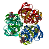
OUR DYNAMIC LIGHT SCATTERING EXPERIMENTS HAVE SHOWN THATTHE PROTEIN EXISTS AS A MONOMER IN SOLUTION, SUGGESTINGTHAT THE HEXAMER DESCRIBED IN REMARK 350 IS AN ARTIFACTOF CRYSTALLIZATION ONLY. THE PROTEIN IS KNOWN TO FUNCTIONAS A MONOMER
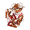
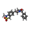
Mass: 17631.648 Da / Num. of mol.: 1 / Fragment: CATALYTIC DOMAIN, RESIDUES 106-264 Source method: isolated from a genetically manipulated source Details: COMPLEXED WITH A SMALL MOLECULE INHIBITOR 2-(1,3-DIOXO-1,3-DIHYDRO-2H-ISOINDOL-2-YL)ETHYL-4-(4-ETHOXY[1,1-BIPHENYL]-4-YL)-4-OXOBUTANOIC ACID FORMULA C19H19N2O4S, AND ALSO WITH ...Details: COMPLEXED WITH A SMALL MOLECULE INHIBITOR 2-(1,3-DIOXO-1,3-DIHYDRO-2H-ISOINDOL-2-YL)ETHYL-4-(4-ETHOXY[1,1-BIPHENYL]-4-YL)-4-OXOBUTANOIC ACID FORMULA C19H19N2O4S, AND ALSO WITH ACETOHYDROXAMIC ACID OR 2-HYDROXYAMINO-2-ETHANAL FORMULA, C2H5NO2 Source: (gene. exp.) HOMO SAPIENS (human) / Plasmid: PGEMEX1 / Production host: ESCHERICHIA COLI (E. coli) / Strain (production host): BL21(DE3) / References: UniProt: P39900, macrophage elastase
Mass: 18.015 Da / Num. of mol.: 97 / Source method: isolated from a natural source / Formula: H2O
-
Details
Compound details
MAY BE INVOLVED IN TISSUE INJURY AND REMODELING. HAS SIGNIFICANT ELASTOLYTIC ACTIVITY. BINDS 2 ZINC ...MAY BE INVOLVED IN TISSUE INJURY AND REMODELING. HAS SIGNIFICANT ELASTOLYTIC ACTIVITY. BINDS 2 ZINC IONS AND 4 CALCIUM IONS PER SUBUNIT. FOUND IN ALVEOLAR MACROPHAGES BUT NOT IN PERIPHERAL BLOOD MONOCYTES.
-
Experimental details
-
Experiment
Experiment
Method: X-RAY DIFFRACTION / Number of used crystals: 1
-
Sample preparation
Crystal
Density Matthews: 4.2 Å3/Da / Density % sol: 71 %
Crystal grow
Temperature: 277 K / Method: vapor diffusion, hanging drop / pH: 6.5 Details: 0.1M SODIUM CACODYLATE PH 6.4, 1.4M AMMONIUM ACETATE, 4 C
In the structure databanks used in Yorodumi, some data are registered as the other names, "COVID-19 virus" and "2019-nCoV". Here are the details of the virus and the list of structure data.
Jan 31, 2019. EMDB accession codes are about to change! (news from PDBe EMDB page)
EMDB accession codes are about to change! (news from PDBe EMDB page)
The allocation of 4 digits for EMDB accession codes will soon come to an end. Whilst these codes will remain in use, new EMDB accession codes will include an additional digit and will expand incrementally as the available range of codes is exhausted. The current 4-digit format prefixed with “EMD-” (i.e. EMD-XXXX) will advance to a 5-digit format (i.e. EMD-XXXXX), and so on. It is currently estimated that the 4-digit codes will be depleted around Spring 2019, at which point the 5-digit format will come into force.
The EM Navigator/Yorodumi systems omit the EMD- prefix.
Related info.:Q: What is EMD? / ID/Accession-code notation in Yorodumi/EM Navigator
Yorodumi is a browser for structure data from EMDB, PDB, SASBDB, etc.
This page is also the successor to EM Navigator detail page, and also detail information page/front-end page for Omokage search.
The word "yorodu" (or yorozu) is an old Japanese word meaning "ten thousand". "mi" (miru) is to see.
Related info.:EMDB / PDB / SASBDB / Comparison of 3 databanks / Yorodumi Search / Aug 31, 2016. New EM Navigator & Yorodumi / Yorodumi Papers / Jmol/JSmol / Function and homology information / Changes in new EM Navigator and Yorodumi
 Movie
Movie Controller

 Yorodumi
 Open data
 Basic information
 Components
 Keywords
 Function and homology information
 HOMO SAPIENS (human)
 X-RAY DIFFRACTION /
  Authors
 Citation
 Structure visualization
 Downloads & links
 Other downloads
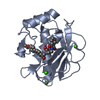


 PDBj
PDBj


 Assembly
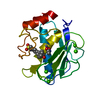


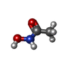


 Mass: 75.067 Da / Num. of mol.: 1 / Source method: obtained synthetically / Formula: C2H5NO2 / Comment: medication*YM
 Mass: 372.438 Da / Num. of mol.: 1 / Source method: obtained synthetically / Formula: C19H20N2O4S
 Mass: 65.409 Da / Num. of mol.: 2 / Source method: obtained synthetically / Formula: Zn
 Mass: 40.078 Da / Num. of mol.: 4 / Source method: obtained synthetically / Formula: Ca
 Sample preparation
 Processing