 Movie
Movie Controller

[English] 日本語
 Yorodumi
- PDB-6k2o: Structural basis of glycan recognition in globally predominant hu... -
+ Open data

- Basic information
| Entry | Database: PDB / ID: 6k2o | |||||||||
|---|---|---|---|---|---|---|---|---|---|---|
| Title | Structural basis of glycan recognition in globally predominant human P[8] rotavirus | |||||||||
 Components Components | Outer capsid protein VP4 | |||||||||
 Keywords Keywords | VIRAL PROTEIN / glycan binding specificity / VP8* structure / mucin core 2 / lacto-N-fucopentaose 1 (LNFP1) | |||||||||
| Function / homology |  Function and homology information Function and homology informationhost cell rough endoplasmic reticulum / permeabilization of host organelle membrane involved in viral entry into host cell / host cytoskeleton / viral outer capsid / host cell endoplasmic reticulum-Golgi intermediate compartment / virion attachment to host cell / host cell plasma membrane Similarity search - Function | |||||||||
| Biological species |  Rotavirus A Rotavirus A | |||||||||
| Method |  X-RAY DIFFRACTION / SYNCHROTRON / MOLECULAR REPLACEMENT / Resolution: 2.296 Å X-RAY DIFFRACTION / SYNCHROTRON / MOLECULAR REPLACEMENT / Resolution: 2.296 Å | |||||||||
 Authors Authors | Duan, Z. / Sun, X. | |||||||||
| Funding support |  China, 1items China, 1items
| |||||||||
 Citation Citation | Journal: Virol Sin / Year: 2020 Title: Structural Basis of Glycan Recognition in Globally Predominant Human P[8] Rotavirus. Authors: Sun, X. / Dang, L. / Li, D. / Qi, J. / Wang, M. / Chai, W. / Zhang, Q. / Wang, H. / Bai, R. / Tan, M. / Duan, Z. | |||||||||
| History |
|
- Structure visualization
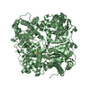
| Structure viewer | Molecule: MolmilJmol/JSmol |
|---|
- Downloads & links
-Download
| PDBx/mmCIF format | 6k2o.cif.gz | 53.6 KB | Display | PDBx/mmCIF format |
|---|---|---|---|---|
| PDB format | pdb6k2o.ent.gz | 35.2 KB | Display | PDB format |
| PDBx/mmJSON format | 6k2o.json.gz | Tree view | PDBx/mmJSON format | |
| Others |  Other downloads Other downloads |
-Validation report
| Arichive directory | https://data.pdbj.org/pub/pdb/validation_reports/k2/6k2oftp://data.pdbj.org/pub/pdb/validation_reports/k2/6k2o | HTTPS FTP |
|---|
-Related structure data
| Related structure data |  6k2nC 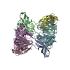 5jdbS S: Starting model for refinement C: citing same article ( |
|---|---|
| Similar structure data |


-Links
 PDBj
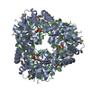
PDBj- Assembly
| Deposited unit | 
| ||||||||
|---|---|---|---|---|---|---|---|---|---|
| 1 | 
| ||||||||
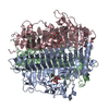
| Unit cell |
|
-Components
| #1: Protein | Mass: 18892.783 Da / Num. of mol.: 1 / Fragment: UNP residues 65-223 Source method: isolated from a genetically manipulated source Source: (gene. exp.) Rotavirus AProduction host: References: UniProt: E2EA82 |
|---|---|
| #2: Polysaccharide | alpha-L-fucopyranose-(1-2)-beta-D-galactopyranose-(1-3)-2-acetamido-2-deoxy-beta-D-glucopyranose-(1- ...alpha-L-fucopyranose-(1-2)-beta-D-galactopyranose-(1-3)-2-acetamido-2-deoxy-beta-D-glucopyranose-(1-3)-beta-D-galactopyranose Source method: isolated from a genetically manipulated source |
| #3: Chemical | ChemComp-NA /   Mass: 22.990 Da / Num. of mol.: 1 Mass: 22.990 Da / Num. of mol.: 1Source method: isolated from a genetically manipulated source Formula: Na / Feature type: SUBJECT OF INVESTIGATION |
| #4: Water | ChemComp-HOH /  Mass: 18.015 Da / Num. of mol.: 80 Mass: 18.015 Da / Num. of mol.: 80Source method: isolated from a genetically manipulated source Formula: H2O |
| Has ligand of interest | Y |
-Experimental details
-Experiment
| Experiment | Method: X-RAY DIFFRACTION / Number of used crystals: 1 |
|---|
- Sample preparation
| Crystal | Density Matthews: 2.88 Å3/Da / Density % sol: 57.33 % |
|---|---|
| Crystal grow | Temperature: 291 K / Method: vapor diffusion, sitting drop Details: 0.1M Sodium acetate trihydrate pH 4.5, 2.0M ammonium sulfate |
-Data collection
| Diffraction | Mean temperature: 113 K / Serial crystal experiment: N |
|---|---|
| Diffraction source | Source: SYNCHROTRON / Site: SSRF / Beamline: BL17U / Wavelength: 0.97852 Å |
| Detector | Type: ADSC QUANTUM 315r / Detector: CCD / Date: Dec 22, 2018 |
| Radiation | Protocol: SINGLE WAVELENGTH / Monochromatic (M) / Laue (L): M / Scattering type: x-ray |
| Radiation wavelength | Wavelength: 0.97852 Å / Relative weight: 1 |
| Reflection | Resolution: 2.296→50 Å / Num. obs: 10510 / % possible obs: 100 % / Redundancy: 25.8 % / Biso Wilson estimate: 29.25 Å2 / Rmerge(I) obs: 0.159 / Rsym value: 0.159 / Net I/σ(I): 22.524 |
| Reflection shell | Resolution: 2.3→2.38 Å / Rmerge(I) obs: 0.86 / Mean I/σ(I) obs: 4.889 / Num. unique obs: 10510 / Rsym value: 0.86 |
- Processing
| Software |
| ||||||||||||||||||
|---|---|---|---|---|---|---|---|---|---|---|---|---|---|---|---|---|---|---|---|
| Refinement | Method to determine structure: MOLECULAR REPLACEMENT Starting model: 5JDB Resolution: 2.296→44.711 Å / Cross valid method: FREE R-VALUE
| ||||||||||||||||||
| Refinement step | Cycle: LAST / Resolution: 2.296→44.711 Å
| ||||||||||||||||||
| LS refinement shell | Resolution: 2.296→2.5 Å
|
