 Movie
Movie Controller

[English] 日本語
 Yorodumi
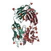
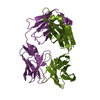
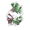
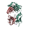
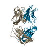
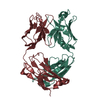
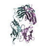
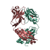
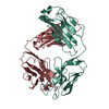
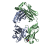
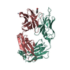
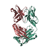
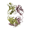
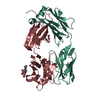
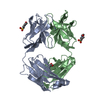
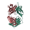
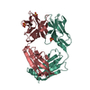
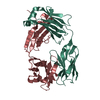
- PDB-5l9d: AFAMIN ANTIBODY FRAGMENT, N14 FAB, L1- GLYCOSYLATED, CRYSTAL FORM... -
+ Open data

- Basic information
| Entry | Database: PDB / ID: 5l9d | ||||||
|---|---|---|---|---|---|---|---|
| Title | AFAMIN ANTIBODY FRAGMENT, N14 FAB, L1- GLYCOSYLATED, CRYSTAL FORM I, parsimonious model | ||||||
 Components Components | (MOUSE ANTIBODY FAB FRAGMENT, IGG1-KAPPA ...) x 2 | ||||||
 Keywords Keywords | IMMUNE SYSTEM / Afamin antibody / Fab fragment / IgG1-kappa / light chain glycosilation | ||||||
| Function / homology | Immunoglobulins / Immunoglobulin-like / Sandwich / Mainly Beta / : / 3,6,9,12,15,18,21-HEPTAOXATRICOSANE-1,23-DIOL / DI(HYDROXYETHYL)ETHER / TRIETHYLENE GLYCOL Function and homology information Function and homology information | ||||||
| Biological species |  | ||||||
| Method |  X-RAY DIFFRACTION / SYNCHROTRON / MOLECULAR REPLACEMENT / Resolution: 1.88 Å X-RAY DIFFRACTION / SYNCHROTRON / MOLECULAR REPLACEMENT / Resolution: 1.88 Å | ||||||
 Authors Authors | Rupp, B. / Naschberger, A. | ||||||
| Funding support |  Austria, 1items Austria, 1items
| ||||||
 Citation Citation | Journal: Acta Crystallogr D Struct Biol / Year: 2016 Title: The N14 anti-afamin antibody Fab: a rare VL1 CDR glycosylation, crystallographic re-sequencing, molecular plasticity and conservative versus enthusiastic modelling. Authors: Naschberger, A. / Furnrohr, B.G. / Lenac Rovis, T. / Malic, S. / Scheffzek, K. / Dieplinger, H. / Rupp, B. | ||||||
| History |
|
- Structure visualization
| Structure viewer | Molecule: MolmilJmol/JSmol |
|---|
- Downloads & links
-Download
| PDBx/mmCIF format | 5l9d.cif.gz | 186.6 KB | Display | PDBx/mmCIF format |
|---|---|---|---|---|
| PDB format | pdb5l9d.ent.gz | 146.7 KB | Display | PDB format |
| PDBx/mmJSON format | 5l9d.json.gz | Tree view | PDBx/mmJSON format | |
| Others |  Other downloads Other downloads |
-Validation report
| Arichive directory | https://data.pdbj.org/pub/pdb/validation_reports/l9/5l9dftp://data.pdbj.org/pub/pdb/validation_reports/l9/5l9d | HTTPS FTP |
|---|
-Related structure data
| Related structure data |  5l7xC 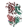 5l88C  5lghC 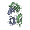 1il1S C: citing same article ( S: Starting model for refinement |
|---|---|
| Similar structure data |


-Links
 PDBj
PDBj

- Assembly
| Deposited unit | 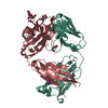
| ||||||||
|---|---|---|---|---|---|---|---|---|---|
| 1 |
| ||||||||
| Unit cell |
|
-Components
-Antibody , 2 types, 2 molecules HL
| #1: Antibody | Mass: 23686.490 Da / Num. of mol.: 1 / Source method: isolated from a natural source / Source: (natural) |
|---|---|
| #2: Antibody | Mass: 23658.104 Da / Num. of mol.: 1 / Source method: isolated from a natural source / Source: (natural) |
-Sugars , 1 types, 1 molecules 
| #8: Sugar | ChemComp-NAG /  Type: D-saccharide, beta linking / Mass: 221.208 Da / Num. of mol.: 1 Type: D-saccharide, beta linking / Mass: 221.208 Da / Num. of mol.: 1Source method: isolated from a genetically manipulated source Formula: C8H15NO6 |
|---|
-Non-polymers , 6 types, 229 molecules 


| #3: Chemical |  Mass: 194.226 Da / Num. of mol.: 2 Mass: 194.226 Da / Num. of mol.: 2Source method: isolated from a genetically manipulated source Formula: C8H18O5 / Comment: precipitant*YM #4: Chemical | ChemComp-PE8 / |  Mass: 370.436 Da / Num. of mol.: 1 Mass: 370.436 Da / Num. of mol.: 1Source method: isolated from a genetically manipulated source Formula: C16H34O9 #5: Chemical | ChemComp-PGE / |  Mass: 150.173 Da / Num. of mol.: 1 Mass: 150.173 Da / Num. of mol.: 1Source method: isolated from a genetically manipulated source Formula: C6H14O4 #6: Chemical |  Mass: 106.120 Da / Num. of mol.: 3 Mass: 106.120 Da / Num. of mol.: 3Source method: isolated from a genetically manipulated source Formula: C4H10O3 #7: Chemical | ChemComp-K / |  Mass: 39.098 Da / Num. of mol.: 1 Mass: 39.098 Da / Num. of mol.: 1Source method: isolated from a genetically manipulated source Formula: K #9: Water | ChemComp-HOH / | Mass: 18.015 Da / Num. of mol.: 221 / Source method: isolated from a natural source / Formula: H2O |
|---|
-Details
| Has protein modification | Y |
|---|
-Experimental details
-Experiment
| Experiment | Method: X-RAY DIFFRACTION / Number of used crystals: 1 |
|---|
- Sample preparation
| Crystal | Density Matthews: 2.19 Å3/Da / Density % sol: 43.9 % Description: BLOCK-SHAPED CRYSTALS WITH SHARP EDGES (0.15 X 0.15 X 0.3 MM) |
|---|---|
| Crystal grow | Temperature: 291 K / Method: vapor diffusion, hanging drop / pH: 5.8 Details: 200NL FAB 10MG/ML IN 20 MM HEPES PH 7.5 REMARK 280 7.5, 150 MM NACL PLUS 200NL 30%W/V PEG 1K, 200MM KF, pH 5.8) Temp details: Air conditioned room |
-Data collection
| Diffraction | Mean temperature: 100 K |
|---|---|
| Diffraction source | Source: SYNCHROTRON / Site: ESRF  / Beamline: ID29 / Wavelength: 1 Å / Beamline: ID29 / Wavelength: 1 Å |
| Detector | Type: DECTRIS PILATUS 6M / Detector: PIXEL / Date: Jun 29, 2015 / Details: TORROIDAL FOCUSSING MIRROR |
| Radiation | Monochromator: Si(111) / Protocol: SINGLE WAVELENGTH / Monochromatic (M) / Laue (L): M / Scattering type: x-ray |
| Radiation wavelength | Wavelength: 1 Å / Relative weight: 1 |
| Reflection | Resolution: 1.88→54.37 Å / Num. obs: 34249 / % possible obs: 99.4 % / Redundancy: 4.5 % / Biso Wilson estimate: 36.6 Å2 / Rmerge(I) obs: 0.076 / Net I/av σ(I): 10 / Net I/σ(I): 99.8 |
| Reflection shell | Resolution: 1.88→1.95 Å / Redundancy: 3.2 % / Rmerge(I) obs: 0.8 / Mean I/σ(I) obs: 1.4 / % possible all: 97.7 |
- Processing
| Software |
| ||||||||||||||||||||||||||||||||||||||||||||||||||||||||||||||||||||||||||||||||||||||||||||||||||||||||||||||||||||||||||||||||||||||||||||||||||||||||||||||||||||||||||||||||||||||
|---|---|---|---|---|---|---|---|---|---|---|---|---|---|---|---|---|---|---|---|---|---|---|---|---|---|---|---|---|---|---|---|---|---|---|---|---|---|---|---|---|---|---|---|---|---|---|---|---|---|---|---|---|---|---|---|---|---|---|---|---|---|---|---|---|---|---|---|---|---|---|---|---|---|---|---|---|---|---|---|---|---|---|---|---|---|---|---|---|---|---|---|---|---|---|---|---|---|---|---|---|---|---|---|---|---|---|---|---|---|---|---|---|---|---|---|---|---|---|---|---|---|---|---|---|---|---|---|---|---|---|---|---|---|---|---|---|---|---|---|---|---|---|---|---|---|---|---|---|---|---|---|---|---|---|---|---|---|---|---|---|---|---|---|---|---|---|---|---|---|---|---|---|---|---|---|---|---|---|---|---|---|---|---|
| Refinement | Method to determine structure: MOLECULAR REPLACEMENT Starting model: 1IL1 Resolution: 1.88→47.38 Å / Cor.coef. Fo:Fc: 0.967 / Cor.coef. Fo:Fc free: 0.956 / SU B: 8.279 / SU ML: 0.119 / Cross valid method: THROUGHOUT / ESU R: 0.155 / ESU R Free: 0.139 / Stereochemistry target values: MAXIMUM LIKELIHOOD / Details: HYDROGENS HAVE BEEN ADDED IN THE RIDING POSITIONS
| ||||||||||||||||||||||||||||||||||||||||||||||||||||||||||||||||||||||||||||||||||||||||||||||||||||||||||||||||||||||||||||||||||||||||||||||||||||||||||||||||||||||||||||||||||||||
| Solvent computation | Ion probe radii: 0.8 Å / Shrinkage radii: 0.8 Å / VDW probe radii: 1.2 Å / Solvent model: MASK | ||||||||||||||||||||||||||||||||||||||||||||||||||||||||||||||||||||||||||||||||||||||||||||||||||||||||||||||||||||||||||||||||||||||||||||||||||||||||||||||||||||||||||||||||||||||
| Displacement parameters | Biso mean: 35.896 Å2
| ||||||||||||||||||||||||||||||||||||||||||||||||||||||||||||||||||||||||||||||||||||||||||||||||||||||||||||||||||||||||||||||||||||||||||||||||||||||||||||||||||||||||||||||||||||||
| Refinement step | Cycle: LAST / Resolution: 1.88→47.38 Å
| ||||||||||||||||||||||||||||||||||||||||||||||||||||||||||||||||||||||||||||||||||||||||||||||||||||||||||||||||||||||||||||||||||||||||||||||||||||||||||||||||||||||||||||||||||||||
| Refine LS restraints |
|