| Software | | Name | Version | Classification |
|---|
| DENZO | | data reduction| SCALEPACK | | data scaling X-PLOR X-PLOR | | model building| CNS | 1 | refinement| X-PLOR | | phasing | | | | |
|
|---|
| Refinement | Method to determine structure: MOLECULAR REPLACEMENT / Resolution: 2.2→19.74 Å / Rfactor Rfree error: 0.006 / Data cutoff high absF: 124012.54 / Data cutoff low absF: 0 / Isotropic thermal model: RESTRAINED / Cross valid method: THROUGHOUT / σ(F): 0 / σ(I): 0
| Rfactor | Num. reflection | % reflection | Selection details |
|---|
| Rfree | 0.246 | 1833 | 9.9 % | RANDOM |
|---|
| Rwork | 0.1883 | - | - | - |
|---|
| all | 0.187 | 29382 | - | - |
|---|
| obs | 0.187 | 18605 | 83.8 % | - |
|---|
|
|---|
| Solvent computation | Solvent model: FLAT MODEL / Bsol: 60.51 Å2 / ksol: 0.345 e/Å3 |
|---|
| Displacement parameters | Biso mean: 35.8 Å2
| Baniso -1 | Baniso -2 | Baniso -3 |
|---|
| 1- | -6.6 Å2 | 0 Å2 | 0 Å2 |
|---|
| 2- | - | 7 Å2 | 0 Å2 |
|---|
| 3- | - | - | -0.4 Å2 |
|---|
|
|---|
| Refine analyze | | Free | Obs |
|---|
| Luzzati coordinate error | 0.33 Å | 0.25 Å |
|---|
| Luzzati d res low | - | 5 Å |
|---|
| Luzzati sigma a | 0.39 Å | 0.31 Å |
|---|
|
|---|
| Refinement step | Cycle: LAST / Resolution: 2.2→19.74 Å
| Protein | Nucleic acid | Ligand | Solvent | Total |
|---|
| Num. atoms | 3379 | 0 | 0 | 260 | 3639 |
|---|
|
|---|
| Refine LS restraints | | Refine-ID | Type | Dev ideal | Dev ideal target |
|---|
| X-RAY DIFFRACTION | c_bond_d| 0.006 | | | X-RAY DIFFRACTION | c_angle_deg| 1.3 | | | X-RAY DIFFRACTION | c_dihedral_angle_d| 27.3 | | | X-RAY DIFFRACTION | c_improper_angle_d| 0.79 | | | X-RAY DIFFRACTION | c_mcbond_it| 3.1 | 2.5 | | X-RAY DIFFRACTION | c_mcangle_it| 4.5 | 3 | | X-RAY DIFFRACTION | c_scbond_it| 6.3 | 3 | | X-RAY DIFFRACTION | c_scangle_it| 8 | 3.5 | | | | | | | | |
|
|---|
| LS refinement shell | Resolution: 2.19→2.33 Å / Rfactor Rfree error: 0.022 / Total num. of bins used: 6
| Rfactor | Num. reflection | % reflection |
|---|
| Rfree | 0.351 | 250 | 9.9 % |
|---|
| Rwork | 0.288 | 2287 | - |
|---|
| obs | - | 2287 | 69 % |
|---|
|
|---|
| Xplor file | | Refine-ID | Serial no | Param file | Topol file |
|---|
| X-RAY DIFFRACTION | 1 | PROTEIN_REP.PARAMPROTEIN.TOP| X-RAY DIFFRACTION | 2 | WATER_REP.PARAM| WATER.TOP | | | |
|
|---|
| Software | *PLUS Name: CNS / Version: 1 / Classification: refinement |
|---|
| Refinement | *PLUS σ(F): 0 / % reflection Rfree: 9.9 % / Rfactor Rfree: 0.247 |
|---|
| Solvent computation | *PLUS |
|---|
| Displacement parameters | *PLUS Biso mean: 35.8 Å2 |
|---|
| Refine LS restraints | *PLUS | Refine-ID | Type | Dev ideal | Dev ideal target |
|---|
| X-RAY DIFFRACTION | c_angle_deg| 1.3 | | | X-RAY DIFFRACTION | c_dihedral_angle_d | | | X-RAY DIFFRACTION | c_dihedral_angle_deg| 27.3 | | | X-RAY DIFFRACTION | c_improper_angle_d | | | X-RAY DIFFRACTION | c_improper_angle_deg| 0.79 | | | X-RAY DIFFRACTION | c_mcbond_it| 3.1 | 2.5 | | X-RAY DIFFRACTION | c_scbond_it| 6.3 | 3 | | X-RAY DIFFRACTION | c_mcangle_it| 4.5 | 3 | | X-RAY DIFFRACTION | c_scangle_it| 8 | 3.5 | | | | | | | | | |
|
|---|
| LS refinement shell | *PLUS Rfactor Rfree: 0.351 / % reflection Rfree: 9.9 % / Rfactor Rwork: 0.288 |
|---|
 Movie
Movie Controller


 Open data
 Basic information
 Components
 Keywords
 Function and homology information
 Authors
 Citation
 Structure visualization
 Downloads & links
 Other downloads
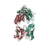


 PDBj
PDBj

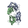
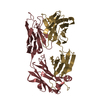
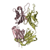
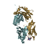
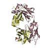
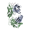
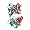
 Assembly
 Mass: 18.015 Da / Num. of mol.: 260 / Source method: isolated from a natural source / Formula: H2O
 Sample preparation
 Processing