 Movie
Movie Controller

[English] 日本語
 Yorodumi
- PDB-5k2x: Crystal structure of M. tuberculosis UspC (tetragonal crystal form) -
+ Open data

- Basic information
| Entry | Database: PDB / ID: 5k2x | ||||||
|---|---|---|---|---|---|---|---|
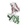
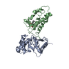
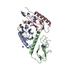
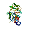
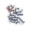
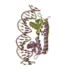
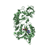
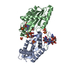
| Title | Crystal structure of M. tuberculosis UspC (tetragonal crystal form) | ||||||
 Components Components | Sugar ABC transporter permease | ||||||
 Keywords Keywords | TRANSPORT PROTEIN / Mycobacterium tuberculosis / ABC transporter / solute binding protein | ||||||
| Function / homology |  Function and homology information Function and homology informationglycerophosphodiester transmembrane transport / glycerol-3-phosphate transmembrane transport / cell envelope / transmembrane transporter activity / membrane Similarity search - Function | ||||||
| Biological species |   Mycobacterium tuberculosis (bacteria) Mycobacterium tuberculosis (bacteria) | ||||||
| Method |  X-RAY DIFFRACTION / SYNCHROTRON / SAD / Resolution: 1.5 Å X-RAY DIFFRACTION / SYNCHROTRON / SAD / Resolution: 1.5 Å | ||||||
 Authors Authors | Futterer, K. / Fullam, E. / Besra, G.S. | ||||||
| Funding support |  United Kingdom, 1items United Kingdom, 1items
| ||||||
 Citation Citation | Journal: Open Biology / Year: 2016 Title: Structural and functional analysis of the solute-binding protein UspC from Mycobacterium tuberculosis that is specific for amino sugars. Authors: Fullam, E. / Prokes, I. / Futterer, K. / Besra, G.S. | ||||||
| History |
|
- Structure visualization
| Structure viewer | Molecule: MolmilJmol/JSmol |
|---|
- Downloads & links
-Download
| PDBx/mmCIF format | 5k2x.cif.gz | 174.7 KB | Display | PDBx/mmCIF format |
|---|---|---|---|---|
| PDB format | pdb5k2x.ent.gz | 135.6 KB | Display | PDB format |
| PDBx/mmJSON format | 5k2x.json.gz | Tree view | PDBx/mmJSON format | |
| Others |  Other downloads Other downloads |
-Validation report
| Arichive directory | https://data.pdbj.org/pub/pdb/validation_reports/k2/5k2xftp://data.pdbj.org/pub/pdb/validation_reports/k2/5k2x | HTTPS FTP |
|---|
-Related structure data


-Links
 PDBj
PDBj


- Assembly
| Deposited unit | 
| ||||||||
|---|---|---|---|---|---|---|---|---|---|
| 1 |
| ||||||||
| Unit cell |
|
-Components
| #1: Protein | Mass: 45227.133 Da / Num. of mol.: 1 Source method: isolated from a genetically manipulated source Details: N-terminally truncated sequence of M. tuberculosis UspC (Rv2318). Source: (gene. exp.) Mycobacterium tuberculosis (bacteria) / Gene: uspC, ERS007688_00434 / Plasmid: pET23b / Production host: | ||||||||
|---|---|---|---|---|---|---|---|---|---|
| #2: Chemical | ChemComp-IOD /   Mass: 126.904 Da / Num. of mol.: 14 / Source method: obtained synthetically / Formula: I Mass: 126.904 Da / Num. of mol.: 14 / Source method: obtained synthetically / Formula: I#3: Chemical | ChemComp-EDO / |   Mass: 62.068 Da / Num. of mol.: 1 / Source method: obtained synthetically / Formula: C2H6O2 Mass: 62.068 Da / Num. of mol.: 1 / Source method: obtained synthetically / Formula: C2H6O2#4: Chemical | ChemComp-MPD / ( |   Mass: 118.174 Da / Num. of mol.: 1 / Source method: obtained synthetically / Formula: C6H14O2 / Comment: precipitant*YM Mass: 118.174 Da / Num. of mol.: 1 / Source method: obtained synthetically / Formula: C6H14O2 / Comment: precipitant*YM#5: Water | ChemComp-HOH / |  Mass: 18.015 Da / Num. of mol.: 463 / Source method: isolated from a natural source / Formula: H2O Mass: 18.015 Da / Num. of mol.: 463 / Source method: isolated from a natural source / Formula: H2OHas protein modification | N | |
-Experimental details
-Experiment
| Experiment | Method: X-RAY DIFFRACTION / Number of used crystals: 1 |
|---|
- Sample preparation
| Crystal | Density Matthews: 2.3 Å3/Da / Density % sol: 46.54 % |
|---|---|
| Crystal grow | Temperature: 292 K / Method: vapor diffusion, sitting drop / pH: 6.5 Details: 0.12 M of 1,6-Hexanediol; 1-Butanol; 1,2-Propanediol; 2-Propanol; 1,4-Butanediol; 1,3-Propanediol; 1 M of imidazole, MES; 25% v/v MPD; 25% PEG 1000; 25% w/v PEG 3350 |
-Data collection
| Diffraction | Mean temperature: 100 K |
|---|---|
| Diffraction source | Source: SYNCHROTRON / Site: Diamond / Beamline: I04-1 / Wavelength: 0.9173 Å |
| Detector | Type: DECTRIS PILATUS 2M / Detector: PIXEL / Date: Jan 2, 2012 |
| Radiation | Protocol: SINGLE WAVELENGTH / Monochromatic (M) / Laue (L): M / Scattering type: x-ray |
| Radiation wavelength | Wavelength: 0.9173 Å / Relative weight: 1 |
| Reflection | Resolution: 1.5→45.31 Å / Num. obs: 64354 / % possible obs: 99.4 % / Observed criterion σ(F): 0 / Redundancy: 6.8 % / Biso Wilson estimate: 12.3 Å2 / Rmerge(I) obs: 0.106 / Rsym value: 0.106 / Net I/σ(I): 13.8 |
| Reflection shell | Resolution: 1.5→1.54 Å / Redundancy: 6.9 % / Rmerge(I) obs: 0.808 / Mean I/σ(I) obs: 3.2 / % possible all: 99 |
- Processing
| Software |
| ||||||||||||||||||||||||||||||||||||||||||||||||||||||||||||
|---|---|---|---|---|---|---|---|---|---|---|---|---|---|---|---|---|---|---|---|---|---|---|---|---|---|---|---|---|---|---|---|---|---|---|---|---|---|---|---|---|---|---|---|---|---|---|---|---|---|---|---|---|---|---|---|---|---|---|---|---|---|
| Refinement | Method to determine structure: SAD / Resolution: 1.5→45.31 Å / Cor.coef. Fo:Fc: 0.964 / Cor.coef. Fo:Fc free: 0.944 / SU B: 2.353 / SU ML: 0.044 / Cross valid method: THROUGHOUT / σ(F): 0 / ESU R: 0.069 / ESU R Free: 0.07 / Stereochemistry target values: MAXIMUM LIKELIHOOD Details: U VALUES : WITH TLS ADDED HYDROGENS HAVE BEEN USED IF PRESENT IN THE INPUT
| ||||||||||||||||||||||||||||||||||||||||||||||||||||||||||||
| Solvent computation | Ion probe radii: 0.8 Å / Shrinkage radii: 0.8 Å / VDW probe radii: 1.2 Å / Solvent model: MASK | ||||||||||||||||||||||||||||||||||||||||||||||||||||||||||||
| Displacement parameters | Biso max: 52.63 Å2 / Biso mean: 14.403 Å2 / Biso min: 4.98 Å2
| ||||||||||||||||||||||||||||||||||||||||||||||||||||||||||||
| Refinement step | Cycle: final / Resolution: 1.5→45.31 Å
| ||||||||||||||||||||||||||||||||||||||||||||||||||||||||||||
| Refine LS restraints |
| ||||||||||||||||||||||||||||||||||||||||||||||||||||||||||||
| LS refinement shell | Resolution: 1.5→1.539 Å / Total num. of bins used: 20
| ||||||||||||||||||||||||||||||||||||||||||||||||||||||||||||
| Refinement TLS params. | Method: refined / Origin x: 30.418 Å / Origin y: 13.507 Å / Origin z: 1.891 Å
|
