 Movie
Movie Controller


+ Open data

- Basic information
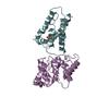
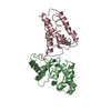
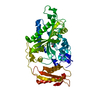
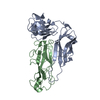
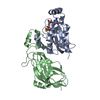
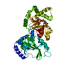
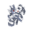
| Entry | Database: PDB / ID: 2zup | ||||||
|---|---|---|---|---|---|---|---|
| Title | Updated crystal structure of DsbB-DsbA complex from E. coli | ||||||
 Components Components |
| ||||||
 Keywords Keywords | OXIDOREDUCTASE / disulfide bond / membrane protein / E. coli / Periplasm / Redox-active center / Cell inner membrane / Cell membrane / Chaperone / Electron transport / Membrane / Transmembrane / Transport | ||||||
| Function / homology |  Function and homology information Function and homology informationoxidoreductase activity, acting on a sulfur group of donors, quinone or similar compound as acceptor / cellular response to antibiotic / protein disulfide isomerase activity / ubiquinone binding / Secretion of toxins / protein-disulfide reductase activity / outer membrane-bounded periplasmic space / response to heat / protein folding / electron transfer activity / plasma membrane Similarity search - Function | ||||||
| Biological species |  | ||||||
| Method |  X-RAY DIFFRACTION / SYNCHROTRON / MIR / Resolution: 3.7 Å X-RAY DIFFRACTION / SYNCHROTRON / MIR / Resolution: 3.7 Å | ||||||
 Authors Authors | Inaba, K. / Suzuki, M. / Murakami, S. / Nakagawa, A. | ||||||
 Citation Citation | Journal: Embo J. / Year: 2009 Title: Dynamic nature of disulphide bond formation catalysts revealed by crystal structures of DsbB Authors: Inaba, K. / Murakami, S. / Nakagawa, A. / Iida, H. / Kinjo, M. / Ito, K. / Suzuki, M. | ||||||
| History |
|
- Structure visualization
| Structure viewer | Molecule: MolmilJmol/JSmol |
|---|
- Downloads & links
-Download
| PDBx/mmCIF format | 2zup.cif.gz | 79.4 KB | Display | PDBx/mmCIF format |
|---|---|---|---|---|
| PDB format | pdb2zup.ent.gz | 59.5 KB | Display | PDB format |
| PDBx/mmJSON format | 2zup.json.gz | Tree view | PDBx/mmJSON format | |
| Others |  Other downloads Other downloads |
-Validation report
| Arichive directory | https://data.pdbj.org/pub/pdb/validation_reports/zu/2zupftp://data.pdbj.org/pub/pdb/validation_reports/zu/2zup | HTTPS FTP |
|---|
-Related structure data
| Related structure data |  2zuqC 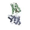 2hi7S S: Starting model for refinement C: citing same article ( |
|---|---|
| Similar structure data |


-Links
 PDBj
PDBj


- Assembly
| Deposited unit | 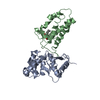
| ||||||||
|---|---|---|---|---|---|---|---|---|---|
| 1 |
| ||||||||
| Unit cell |
| ||||||||
| Components on special symmetry positions |
|
-Components
| #1: Protein | Mass: 21122.959 Da / Num. of mol.: 1 / Mutation: C33A Source method: isolated from a genetically manipulated source Source: (gene. exp.) References: UniProt: P0AEG4, Oxidoreductases; Acting on a sulfur group of donors; With a disulfide as acceptor |
|---|---|
| #2: Protein | Mass: 20106.982 Da / Num. of mol.: 1 / Mutation: C8A,C49V,C130S Source method: isolated from a genetically manipulated source Source: (gene. exp.) References: UniProt: P0A6M2, Oxidoreductases; Acting on a sulfur group of donors; With a quinone or similar compound as acceptor |
| #3: Chemical | ChemComp-ZN /   Mass: 65.409 Da / Num. of mol.: 1 / Source method: obtained synthetically / Formula: Zn Mass: 65.409 Da / Num. of mol.: 1 / Source method: obtained synthetically / Formula: Zn |
| #4: Chemical | ChemComp-UQ1 / 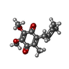  Mass: 250.290 Da / Num. of mol.: 1 / Source method: obtained synthetically / Formula: C14H18O4 Mass: 250.290 Da / Num. of mol.: 1 / Source method: obtained synthetically / Formula: C14H18O4 |
| Has protein modification | Y |
-Experimental details
-Experiment
| Experiment | Method: X-RAY DIFFRACTION / Number of used crystals: 1 |
|---|
- Sample preparation
| Crystal | Density Matthews: 5.474029 Å3/Da / Density % sol: 77.530258 % |
|---|---|
| Crystal grow | Temperature: 293 K / Method: evaporation / pH: 7 Details: 23% Jeffamine ED2001, 80mM HEPES, 14.4% glycerol, 2mM ZnCl2, pH 7.0, EVAPORATION, temperature 293K |
-Data collection
| Diffraction | Mean temperature: 100 K |
|---|---|
| Diffraction source | Source: SYNCHROTRON / Site: SPring-8  / Beamline: BL44XU / Wavelength: 0.9 Å / Beamline: BL44XU / Wavelength: 0.9 Å |
| Detector | Type: MACSCIENCE / Detector: IMAGE PLATE / Date: Sep 22, 2005 |
| Radiation | Monochromator: SI(111) double crystal / Protocol: SINGLE WAVELENGTH / Monochromatic (M) / Laue (L): M / Scattering type: x-ray |
| Radiation wavelength | Wavelength: 0.9 Å / Relative weight: 1 |
| Reflection | Resolution: 3.7→61.2 Å / Num. obs: 10179 / % possible obs: 99.94 % / Observed criterion σ(I): -4 / Redundancy: 6.9 % / Rmerge(I) obs: 0.06 / Net I/σ(I): 18.7 |
| Reflection shell | Resolution: 3.7→3.9 Å / Redundancy: 7.1 % / Rmerge(I) obs: 0.527 / Mean I/σ(I) obs: 3.8 / Num. unique all: 1450 / % possible all: 99.8 |
- Processing
| Software |
| |||||||||||||||||||||||||||||||||||||||||||||||||||||||||||||||||
|---|---|---|---|---|---|---|---|---|---|---|---|---|---|---|---|---|---|---|---|---|---|---|---|---|---|---|---|---|---|---|---|---|---|---|---|---|---|---|---|---|---|---|---|---|---|---|---|---|---|---|---|---|---|---|---|---|---|---|---|---|---|---|---|---|---|---|
| Refinement | Method to determine structure: MIR Starting model: 2HI7 Resolution: 3.7→20 Å / Cor.coef. Fo:Fc: 0.909 / Cor.coef. Fo:Fc free: 0.928 / SU B: 47.802 / SU ML: 0.688 / Cross valid method: THROUGHOUT / ESU R Free: 0.662 / Stereochemistry target values: MAXIMUM LIKELIHOOD / Details: HYDROGENS HAVE BEEN ADDED IN THE RIDING POSITIONS
| |||||||||||||||||||||||||||||||||||||||||||||||||||||||||||||||||
| Solvent computation | Ion probe radii: 0.8 Å / Shrinkage radii: 0.8 Å / VDW probe radii: 1.4 Å / Solvent model: MASK | |||||||||||||||||||||||||||||||||||||||||||||||||||||||||||||||||
| Displacement parameters | Biso mean: 205.362 Å2
| |||||||||||||||||||||||||||||||||||||||||||||||||||||||||||||||||
| Refinement step | Cycle: LAST / Resolution: 3.7→20 Å
| |||||||||||||||||||||||||||||||||||||||||||||||||||||||||||||||||
| Refine LS restraints |
| |||||||||||||||||||||||||||||||||||||||||||||||||||||||||||||||||
| LS refinement shell | Resolution: 3.7→4.039 Å / Total num. of bins used: 6
|
