 Movie
Movie Controller

[English] 日本語
 Yorodumi
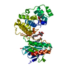
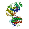
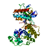
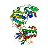
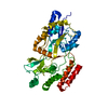
- PDB-5k2y: Crystal structure of M. tuberculosis UspC (monoclinic crystal form) -
+ Open data

- Basic information
| Entry | Database: PDB / ID: 5k2y | ||||||
|---|---|---|---|---|---|---|---|
| Title | Crystal structure of M. tuberculosis UspC (monoclinic crystal form) | ||||||
 Components Components | Probable periplasmic sugar-binding lipoprotein UspC | ||||||
 Keywords Keywords | TRANSPORT PROTEIN / Mycobacterium tuberculosis / ABC transporter / solute binding protein | ||||||
| Function / homology |  Function and homology information Function and homology informationglycerophosphodiester transmembrane transport / glycerol-3-phosphate transmembrane transport / cell envelope / transmembrane transporter activity / membrane Similarity search - Function | ||||||
| Biological species |   Mycobacterium tuberculosis (bacteria) Mycobacterium tuberculosis (bacteria) | ||||||
| Method |  X-RAY DIFFRACTION / SYNCHROTRON / SAD / Resolution: 2.41 Å X-RAY DIFFRACTION / SYNCHROTRON / SAD / Resolution: 2.41 Å | ||||||
 Authors Authors | Futterer, K. / Fullam, E. / Besra, G.S. | ||||||
| Funding support |  United Kingdom, 1items United Kingdom, 1items
| ||||||
 Citation Citation | Journal: Open Biology / Year: 2016 Title: Structural and functional analysis of the solute-binding protein UspC from Mycobacterium tuberculosis that is specific for amino sugars. Authors: Fullam, E. / Prokes, I. / Futterer, K. / Besra, G.S. | ||||||
| History |
|
- Structure visualization
| Structure viewer | Molecule: MolmilJmol/JSmol |
|---|
- Downloads & links
-Download
| PDBx/mmCIF format | 5k2y.cif.gz | 297.4 KB | Display | PDBx/mmCIF format |
|---|---|---|---|---|
| PDB format | pdb5k2y.ent.gz | 241.1 KB | Display | PDB format |
| PDBx/mmJSON format | 5k2y.json.gz | Tree view | PDBx/mmJSON format | |
| Others |  Other downloads Other downloads |
-Validation report
| Arichive directory | https://data.pdbj.org/pub/pdb/validation_reports/k2/5k2yftp://data.pdbj.org/pub/pdb/validation_reports/k2/5k2y | HTTPS FTP |
|---|
-Related structure data


-Links
 PDBj
PDBj


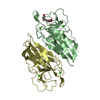
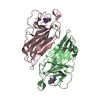
- Assembly
| Deposited unit | 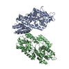
| ||||||||||||||||||
|---|---|---|---|---|---|---|---|---|---|---|---|---|---|---|---|---|---|---|---|
| 1 | 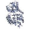
| ||||||||||||||||||
| 2 | 
| ||||||||||||||||||
| Unit cell |
| ||||||||||||||||||
| Noncrystallographic symmetry (NCS) | NCS domain:
NCS domain segments: Component-ID: _ / Ens-ID: 1 / Beg auth comp-ID: ILE / Beg label comp-ID: ILE / End auth comp-ID: THR / End label comp-ID: THR / Refine code: _ / Auth seq-ID: 39 - 438 / Label seq-ID: 1 - 400
|
-Components
| #1: Protein | Mass: 44729.539 Da / Num. of mol.: 2 Source method: isolated from a genetically manipulated source Details: N-terminally truncated UspC from Mycobacterium tuberculosis with C-terminal hexa-histidine tag from pET23b plasmid Source: (gene. exp.) Mycobacterium tuberculosis (bacteria) / Gene: uspC, Rv2318 / Plasmid: pET23b / Production host: #2: Water | ChemComp-HOH / |  Mass: 18.015 Da / Num. of mol.: 52 / Source method: isolated from a natural source / Formula: H2O Mass: 18.015 Da / Num. of mol.: 52 / Source method: isolated from a natural source / Formula: H2O |
|---|
-Experimental details
-Experiment
| Experiment | Method: X-RAY DIFFRACTION / Number of used crystals: 1 |
|---|
- Sample preparation
| Crystal | Density Matthews: 2.33 Å3/Da / Density % sol: 47.24 % |
|---|---|
| Crystal grow | Temperature: 292 K / Method: vapor diffusion, sitting drop / pH: 6.5 Details: Precipitant mix: 25% v/v MPD; 25% PEG 1000; 25% w/v PEG 3350 Halogen mix: 0.3M Sodium fluoride; 0.3M Sodium bromide; 0.3M Sodium iodide Buffer: 1.0 M Imidazole; MES monohydrate (acid) |
-Data collection
| Diffraction | Mean temperature: 100 K |
|---|---|
| Diffraction source | Source: SYNCHROTRON / Site: Diamond / Beamline: I04 / Wavelength: 0.9795 Å |
| Detector | Type: DECTRIS PILATUS 2M / Detector: PIXEL / Date: Oct 23, 2011 |
| Radiation | Protocol: SINGLE WAVELENGTH / Monochromatic (M) / Laue (L): M / Scattering type: x-ray |
| Radiation wavelength | Wavelength: 0.9795 Å / Relative weight: 1 |
| Reflection | Resolution: 2.41→87.9 Å / Num. obs: 30838 / % possible obs: 99.6 % / Observed criterion σ(F): 0 / Redundancy: 7.3 % / Biso Wilson estimate: 38.9 Å2 / Rmerge(I) obs: 0.113 / Rsym value: 0.113 / Net I/σ(I): 14.3 |
| Reflection shell | Resolution: 2.41→2.47 Å / Redundancy: 6 % / Rmerge(I) obs: 0.664 / Mean I/σ(I) obs: 2.3 / % possible all: 95.2 |
- Processing
| Software |
| |||||||||||||||||||||||||||||||||||||||||||||||||||||||||||||||||||||||||||
|---|---|---|---|---|---|---|---|---|---|---|---|---|---|---|---|---|---|---|---|---|---|---|---|---|---|---|---|---|---|---|---|---|---|---|---|---|---|---|---|---|---|---|---|---|---|---|---|---|---|---|---|---|---|---|---|---|---|---|---|---|---|---|---|---|---|---|---|---|---|---|---|---|---|---|---|---|
| Refinement | Method to determine structure: SAD / Resolution: 2.41→87.88 Å / Cor.coef. Fo:Fc: 0.934 / Cor.coef. Fo:Fc free: 0.888 / SU B: 22.395 / SU ML: 0.234 / Cross valid method: THROUGHOUT / σ(F): 0 / ESU R: 0.514 / ESU R Free: 0.288 Details: U VALUES : WITH TLS ADDED HYDROGENS HAVE BEEN USED IF PRESENT IN THE INPUT
| |||||||||||||||||||||||||||||||||||||||||||||||||||||||||||||||||||||||||||
| Solvent computation | Ion probe radii: 0.8 Å / Shrinkage radii: 0.8 Å / VDW probe radii: 1.2 Å | |||||||||||||||||||||||||||||||||||||||||||||||||||||||||||||||||||||||||||
| Displacement parameters | Biso max: 84.68 Å2 / Biso mean: 34.667 Å2 / Biso min: 7.98 Å2
| |||||||||||||||||||||||||||||||||||||||||||||||||||||||||||||||||||||||||||
| Refinement step | Cycle: final / Resolution: 2.41→87.88 Å
| |||||||||||||||||||||||||||||||||||||||||||||||||||||||||||||||||||||||||||
| Refine LS restraints |
| |||||||||||||||||||||||||||||||||||||||||||||||||||||||||||||||||||||||||||
| Refine LS restraints NCS | Ens-ID: 1 / Number: 994 / Refine-ID: X-RAY DIFFRACTION / Type: interatomic distance / Rms dev position: 0.15 Å / Weight position: 0.05
| |||||||||||||||||||||||||||||||||||||||||||||||||||||||||||||||||||||||||||
| LS refinement shell | Resolution: 2.41→2.473 Å / Total num. of bins used: 20
| |||||||||||||||||||||||||||||||||||||||||||||||||||||||||||||||||||||||||||
| Refinement TLS params. | Method: refined / Refine-ID: X-RAY DIFFRACTION
| |||||||||||||||||||||||||||||||||||||||||||||||||||||||||||||||||||||||||||
| Refinement TLS group |
|
