 Movie
Movie Controller

[English] 日本語
 Yorodumi
- PDB-6tts: Crystal structure of the GGDEF domain of DgcB from Caulobacter cr... -
+ Open data

- Basic information
| Entry | Database: PDB / ID: 6tts | ||||||
|---|---|---|---|---|---|---|---|
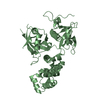
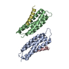
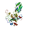
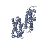
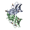
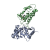
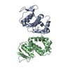
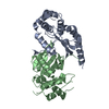
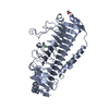
| Title | Crystal structure of the GGDEF domain of DgcB from Caulobacter crescentus in complex with c-di-GMP | ||||||
 Components Components | GGDEF diguanylate cyclase DgcB | ||||||
 Keywords Keywords | SIGNALING PROTEIN / GGDEF / c-di-GMP / cyclic di-GMP / Caulobacter / DgcB / diguanylate cyclase / DGC | ||||||
| Function / homology |  Function and homology information Function and homology informationnegative regulation of bacterial-type flagellum-dependent cell motility / diguanylate cyclase / diguanylate cyclase activity / cell adhesion involved in single-species biofilm formation / nucleotide binding / plasma membrane Similarity search - Function | ||||||
| Biological species |  Caulobacter vibrioides (bacteria) Caulobacter vibrioides (bacteria) | ||||||
| Method |  X-RAY DIFFRACTION / SYNCHROTRON / MOLECULAR REPLACEMENT / Resolution: 2.5 Å X-RAY DIFFRACTION / SYNCHROTRON / MOLECULAR REPLACEMENT / Resolution: 2.5 Å | ||||||
 Authors Authors | Holzschuh, F. / Schirmer, T. / Teixeira, R. | ||||||
| Funding support |  Switzerland, 1items Switzerland, 1items
| ||||||
 Citation Citation | Journal: To Be Published Title: Crystal structure of the GGDEF domain of DgcB from Caulobacter crescentus in complex with c-di-GMP Authors: Holzschuh, F. / Schirmer, T. / Teixeira, R. | ||||||
| History |
|
- Structure visualization
| Structure viewer | Molecule: MolmilJmol/JSmol |
|---|
- Downloads & links
-Download
| PDBx/mmCIF format | 6tts.cif.gz | 98.5 KB | Display | PDBx/mmCIF format |
|---|---|---|---|---|
| PDB format | pdb6tts.ent.gz | 65.6 KB | Display | PDB format |
| PDBx/mmJSON format | 6tts.json.gz | Tree view | PDBx/mmJSON format | |
| Others |  Other downloads Other downloads |
-Validation report
| Arichive directory | https://data.pdbj.org/pub/pdb/validation_reports/tt/6ttsftp://data.pdbj.org/pub/pdb/validation_reports/tt/6tts | HTTPS FTP |
|---|
-Related structure data
| Related structure data |  3ezuS  3hvaS  3i5cS  3iclS  3ignS  3qyyS 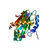 3tvkS 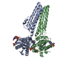 4h54S  4iobS  4urgS 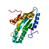 4wxwS  4ymeS 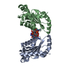 4zmmS 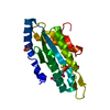 4zvfS  5euhS S: Starting model for refinement |
|---|---|
| Similar structure data |

-Links
 PDBj
PDBj


- Assembly
| Deposited unit | 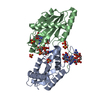
| ||||||||||||
|---|---|---|---|---|---|---|---|---|---|---|---|---|---|
| 1 |
| ||||||||||||
| Unit cell |
|
-Components
| #1: Protein | Mass: 21449.895 Da / Num. of mol.: 2 Source method: isolated from a genetically manipulated source Source: (gene. exp.) Caulobacter vibrioides (strain NA1000 / CB15N) (bacteria)Gene: dgcB, CCNA_01926 / Production host: #2: Chemical | ChemComp-C2E /   Mass: 690.411 Da / Num. of mol.: 5 / Source method: obtained synthetically / Formula: C20H24N10O14P2 Mass: 690.411 Da / Num. of mol.: 5 / Source method: obtained synthetically / Formula: C20H24N10O14P2#3: Chemical | ChemComp-SO4 /   Mass: 96.063 Da / Num. of mol.: 6 / Source method: isolated from a natural source / Formula: SO4 Mass: 96.063 Da / Num. of mol.: 6 / Source method: isolated from a natural source / Formula: SO4#4: Water | ChemComp-HOH / |  Mass: 18.015 Da / Num. of mol.: 16 / Source method: isolated from a natural source / Formula: H2O Mass: 18.015 Da / Num. of mol.: 16 / Source method: isolated from a natural source / Formula: H2OHas ligand of interest | N | |
|---|
-Experimental details
-Experiment
| Experiment | Method: X-RAY DIFFRACTION / Number of used crystals: 1 |
|---|
- Sample preparation
| Crystal | Density Matthews: 2.99 Å3/Da / Density % sol: 58.88 % |
|---|---|
| Crystal grow | Temperature: 293 K / Method: vapor diffusion, sitting drop / pH: 5.5 Details: 0.2 M LiSO4, 0.1 M NaAcet pH 5.5, 8% w/v PEG 20k, 8% v/v PEG 500 MME, 500 uM c-di-GMP, 1 mM GTP |
-Data collection
| Diffraction | Mean temperature: 100 K / Serial crystal experiment: N |
|---|---|
| Diffraction source | Source: SYNCHROTRON / Site: SLS / Beamline: X06SA / Wavelength: 0.97947 Å |
| Detector | Type: DECTRIS EIGER X 16M / Detector: PIXEL / Date: Jun 10, 2018 |
| Radiation | Protocol: SINGLE WAVELENGTH / Monochromatic (M) / Laue (L): M / Scattering type: x-ray |
| Radiation wavelength | Wavelength: 0.97947 Å / Relative weight: 1 |
| Reflection | Resolution: 2.5→64.5 Å / Num. obs: 19045 / % possible obs: 99.9 % / Redundancy: 7.2 % / Biso Wilson estimate: 29.29 Å2 / CC1/2: 0.813 / Rmerge(I) obs: 0.159 / Rpim(I) all: 0.088 / Rrim(I) all: 0.183 / Χ2: 0.95 / Net I/σ(I): 8.3 |
| Reflection shell | Resolution: 2.5→2.6 Å / Redundancy: 3.7 % / Rmerge(I) obs: 0.515 / Mean I/σ(I) obs: 2.3 / Num. unique obs: 2068 / CC1/2: 0.693 / Rpim(I) all: 0.427 / Rrim(I) all: 0.672 / Χ2: 0.89 / % possible all: 99 |
- Processing
| Software |
| ||||||||||||||||||||||||||||||||||||||||||||||||||||||||
|---|---|---|---|---|---|---|---|---|---|---|---|---|---|---|---|---|---|---|---|---|---|---|---|---|---|---|---|---|---|---|---|---|---|---|---|---|---|---|---|---|---|---|---|---|---|---|---|---|---|---|---|---|---|---|---|---|---|
| Refinement | Method to determine structure: MOLECULAR REPLACEMENT Starting model: 3ign, 3hva, 3qyy, 3i5c, 4wxw, 3tvk, 4urg, 4zvf, 4h54, 4zmm, 3icl, 4iob, 5euh, 3ezu, 4yme Resolution: 2.5→50.76 Å / SU ML: 0.3133 / Cross valid method: FREE R-VALUE / σ(F): 1.34 / Phase error: 24.2429 / Stereochemistry target values: GeoStd + Monomer Library
| ||||||||||||||||||||||||||||||||||||||||||||||||||||||||
| Solvent computation | Shrinkage radii: 0.9 Å / VDW probe radii: 1.11 Å / Solvent model: FLAT BULK SOLVENT MODEL | ||||||||||||||||||||||||||||||||||||||||||||||||||||||||
| Displacement parameters | Biso mean: 29.68 Å2 | ||||||||||||||||||||||||||||||||||||||||||||||||||||||||
| Refinement step | Cycle: LAST / Resolution: 2.5→50.76 Å
| ||||||||||||||||||||||||||||||||||||||||||||||||||||||||
| Refine LS restraints |
| ||||||||||||||||||||||||||||||||||||||||||||||||||||||||
| LS refinement shell |
|
