 Movie
Movie Controller


+ Open data

- Basic information
| Entry | Database: PDB / ID: 5kbc | |||||||||
|---|---|---|---|---|---|---|---|---|---|---|
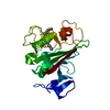
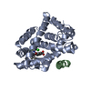
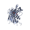
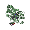
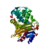
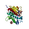
| Title | Crystal structure of Chlamydia trachomatis DsbA | |||||||||
 Components Components | DsbA | |||||||||
 Keywords Keywords | OXIDOREDUCTASE / thioredoxin fold | |||||||||
| Function / homology | Thioredoxin / Thioredoxin-like fold / Thioredoxin-like superfamily / membrane / DsbA Function and homology information Function and homology information | |||||||||
| Biological species |  Chlamydia trachomatis serovar A (bacteria) Chlamydia trachomatis serovar A (bacteria) | |||||||||
| Method |  X-RAY DIFFRACTION / MOLECULAR REPLACEMENT / molecular replacement / Resolution: 2.706 Å X-RAY DIFFRACTION / MOLECULAR REPLACEMENT / molecular replacement / Resolution: 2.706 Å | |||||||||
 Authors Authors | McMahon, R.M. / Groftehauge, M.K. / Martin, J.L. | |||||||||
| Funding support |  Australia, 2items Australia, 2items
| |||||||||
 Citation Citation | Journal: PLoS ONE / Year: 2016 Title: Structural and Biochemical Characterization of Chlamydia trachomatis DsbA Reveals a Cysteine-Rich and Weakly Oxidising Oxidoreductase. Authors: Christensen, S. / Grftehauge, M.K. / Byriel, K. / Huston, W.M. / Furlong, E. / Heras, B. / Martin, J.L. / McMahon, R.M. | |||||||||
| History |
|
- Structure visualization
| Structure viewer | Molecule: MolmilJmol/JSmol |
|---|
- Downloads & links
-Download
| PDBx/mmCIF format | 5kbc.cif.gz | 152.2 KB | Display | PDBx/mmCIF format |
|---|---|---|---|---|
| PDB format | pdb5kbc.ent.gz | 122.4 KB | Display | PDB format |
| PDBx/mmJSON format | 5kbc.json.gz | Tree view | PDBx/mmJSON format | |
| Others |  Other downloads Other downloads |
-Validation report
| Arichive directory | https://data.pdbj.org/pub/pdb/validation_reports/kb/5kbcftp://data.pdbj.org/pub/pdb/validation_reports/kb/5kbc | HTTPS FTP |
|---|
-Related structure data
| Related structure data |  3eu3S S: Starting model for refinement |
|---|---|
| Similar structure data |


-Links
 PDBj
PDBj- Assembly
| Deposited unit | 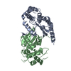
| ||||||||
|---|---|---|---|---|---|---|---|---|---|
| 1 | 
| ||||||||
| 2 | 
| ||||||||
| Unit cell |
|
-Components
| #1: Protein | Mass: 22784.164 Da / Num. of mol.: 2 / Fragment: UNP residues 78-277 Source method: isolated from a genetically manipulated source Source: (gene. exp.) Chlamydia trachomatis serovar A (bacteria)Strain: A2497 / Gene: CTO_0193 / Plasmid: pET21a / Production host: #2: Water | ChemComp-HOH / |  Mass: 18.015 Da / Num. of mol.: 22 / Source method: isolated from a natural source / Formula: H2O Mass: 18.015 Da / Num. of mol.: 22 / Source method: isolated from a natural source / Formula: H2OHas protein modification | Y | |
|---|
-Experimental details
-Experiment
| Experiment | Method: X-RAY DIFFRACTION / Number of used crystals: 1 |
|---|
- Sample preparation
| Crystal | Density Matthews: 2.28 Å3/Da / Density % sol: 46.03 % |
|---|---|
| Crystal grow | Temperature: 298 K / Method: vapor diffusion / pH: 9 Details: 100 mM Bis Tris pH 9.0, 3.1 M sodium formate, 4 % (v/v) Tacsimate |
-Data collection
| Diffraction | Mean temperature: 100 K |
|---|---|
| Diffraction source | Source: ROTATING ANODE / Type: RIGAKU FR-E SUPERBRIGHT / Wavelength: 1.54187 Å |
| Detector | Type: RIGAKU SATURN 944 / Detector: CCD / Date: Aug 24, 2010 |
| Radiation | Protocol: SINGLE WAVELENGTH / Monochromatic (M) / Laue (L): M / Scattering type: x-ray |
| Radiation wavelength | Wavelength: 1.54187 Å / Relative weight: 1 |
| Reflection | Resolution: 2.706→43.319 Å / Num. obs: 11646 / % possible obs: 100 % / Redundancy: 7.1 % / CC1/2: 0.998 / Rmerge(I) obs: 0.101 / Net I/σ(I): 19.6 |
| Reflection shell | Resolution: 2.706→2.715 Å / Redundancy: 7.2 % / Rmerge(I) obs: 0.401 / Mean I/σ(I) obs: 5.9 / % possible all: 86.4 |
-Phasing
| Phasing | Method: molecular replacement | |||||||||
|---|---|---|---|---|---|---|---|---|---|---|
| Phasing MR |
|
- Processing
| Software |
| |||||||||||||||||||||||||||||||||||
|---|---|---|---|---|---|---|---|---|---|---|---|---|---|---|---|---|---|---|---|---|---|---|---|---|---|---|---|---|---|---|---|---|---|---|---|---|
| Refinement | Method to determine structure: MOLECULAR REPLACEMENT Starting model: 3EU3 Resolution: 2.706→43.319 Å / SU ML: 0.42 / Cross valid method: FREE R-VALUE / σ(F): 1.36 / Phase error: 27.5 / Stereochemistry target values: ML
| |||||||||||||||||||||||||||||||||||
| Solvent computation | Shrinkage radii: 0.9 Å / VDW probe radii: 1.11 Å / Solvent model: FLAT BULK SOLVENT MODEL | |||||||||||||||||||||||||||||||||||
| Displacement parameters | Biso max: 95.52 Å2 / Biso mean: 50.488 Å2 / Biso min: 20 Å2 | |||||||||||||||||||||||||||||||||||
| Refinement step | Cycle: final / Resolution: 2.706→43.319 Å
| |||||||||||||||||||||||||||||||||||
| Refine LS restraints |
| |||||||||||||||||||||||||||||||||||
| LS refinement shell | Refine-ID: X-RAY DIFFRACTION / Total num. of bins used: 4
|
