 Movie
Movie Controller

[English] 日本語
 Yorodumi
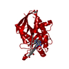
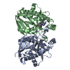
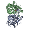
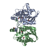
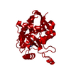
- PDB-4ojt: Helicobacter pylori MTAN complexed with S-ribosylhomocysteine and... -
+ Open data

- Basic information
| Entry | Database: PDB / ID: 4ojt | |||||||||
|---|---|---|---|---|---|---|---|---|---|---|
| Title | Helicobacter pylori MTAN complexed with S-ribosylhomocysteine and adenine | |||||||||
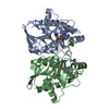
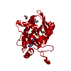
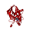
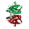
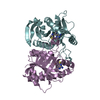
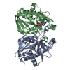
 Components Components | MTA/SAH nucleosidase | |||||||||
 Keywords Keywords | HYDROLASE / Homodimer | |||||||||
| Function / homology |  Function and homology information Function and homology informationaminodeoxyfutalosine nucleosidase / 6-amino-6-deoxyfutalosine hydrolase activity / adenosylhomocysteine nucleosidase / adenosylhomocysteine nucleosidase activity / methylthioadenosine nucleosidase activity / : / nucleoside catabolic process / menaquinone biosynthetic process / : / cytosol Similarity search - Function | |||||||||
| Biological species |   Helicobacter pylori (bacteria) Helicobacter pylori (bacteria) | |||||||||
| Method |  X-RAY DIFFRACTION / SYNCHROTRON / MOLECULAR REPLACEMENT / Resolution: 1.5 Å X-RAY DIFFRACTION / SYNCHROTRON / MOLECULAR REPLACEMENT / Resolution: 1.5 Å | |||||||||
 Authors Authors | Mishra, V. / Ronning, D.R. | |||||||||
 Citation Citation | Journal: Biochemistry / Year: 2012 Title: Crystal structures of the Helicobacter pylori MTAN enzyme reveal specific interactions between S-adenosylhomocysteine and the 5'-alkylthio binding subsite. Authors: Mishra, V. / Ronning, D.R. | |||||||||
| History |
|
- Structure visualization
| Structure viewer | Molecule: MolmilJmol/JSmol |
|---|
- Downloads & links
-Download
| PDBx/mmCIF format | 4ojt.cif.gz | 62.2 KB | Display | PDBx/mmCIF format |
|---|---|---|---|---|
| PDB format | pdb4ojt.ent.gz | 45.4 KB | Display | PDB format |
| PDBx/mmJSON format | 4ojt.json.gz | Tree view | PDBx/mmJSON format | |
| Others |  Other downloads Other downloads |
-Validation report
| Arichive directory | https://data.pdbj.org/pub/pdb/validation_reports/oj/4ojtftp://data.pdbj.org/pub/pdb/validation_reports/oj/4ojt | HTTPS FTP |
|---|
-Related structure data


-Links
 PDBj
PDBj


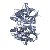
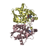
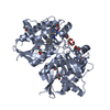
- Assembly
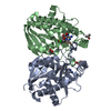
| Deposited unit | 
| ||||||||
|---|---|---|---|---|---|---|---|---|---|
| 1 | 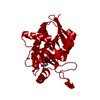
| ||||||||
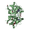
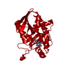
| Unit cell |
| ||||||||
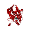
| Components on special symmetry positions |
|
-Components
| #1: Protein | Mass: 25106.951 Da / Num. of mol.: 1 / Fragment: UNP residues 2-230 Source method: isolated from a genetically manipulated source Source: (gene. exp.) Helicobacter pylori (bacteria) / Strain: J99 / ATCC 700824 / Gene: jhp_0082, mtn, mtnN, pfs / Plasmid: pET32 / Production host: References: UniProt: Q9ZMY2, adenosylhomocysteine nucleosidase |
|---|---|
| #2: Sugar | ChemComp-2WP / 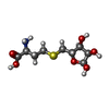  Type: D-saccharide, alpha linking / Mass: 267.299 Da / Num. of mol.: 1 / Source method: obtained synthetically / Formula: C9H17NO6S Type: D-saccharide, alpha linking / Mass: 267.299 Da / Num. of mol.: 1 / Source method: obtained synthetically / Formula: C9H17NO6S |
| #3: Chemical | ChemComp-ADE /   Mass: 135.127 Da / Num. of mol.: 1 / Source method: obtained synthetically / Formula: C5H5N5 Mass: 135.127 Da / Num. of mol.: 1 / Source method: obtained synthetically / Formula: C5H5N5 |
| #4: Water | ChemComp-HOH /  Mass: 18.015 Da / Num. of mol.: 220 / Source method: isolated from a natural source / Formula: H2O Mass: 18.015 Da / Num. of mol.: 220 / Source method: isolated from a natural source / Formula: H2O |
-Experimental details
-Experiment
| Experiment | Method: X-RAY DIFFRACTION / Number of used crystals: 1 |
|---|
- Sample preparation
| Crystal | Density Matthews: 2.56 Å3/Da / Density % sol: 51.94 % |
|---|---|
| Crystal grow | Temperature: 291 K / Method: vapor diffusion, hanging drop / pH: 7.5 Details: 0.05 M magnesium chloride hexahydrate, 0.1 M HEPES, 30 % (v/v) PEG-MME 550, pH 7.5, VAPOR DIFFUSION, HANGING DROP, temperature 291K |
-Data collection
| Diffraction | Mean temperature: 100 K |
|---|---|
| Diffraction source | Source: SYNCHROTRON / Site: APS  / Beamline: 21-ID-D / Wavelength: 0.97856 Å / Beamline: 21-ID-D / Wavelength: 0.97856 Å |
| Detector | Type: MARMOSAIC 300 mm CCD / Detector: CCD / Date: Oct 29, 2011 |
| Radiation | Monochromator: diamond / Protocol: SINGLE WAVELENGTH / Monochromatic (M) / Laue (L): M / Scattering type: x-ray |
| Radiation wavelength | Wavelength: 0.97856 Å / Relative weight: 1 |
| Reflection | Resolution: 1.5→50 Å / Num. obs: 40628 / % possible obs: 98.5 % / Observed criterion σ(F): -3 / Observed criterion σ(I): 0 / Rmerge(I) obs: 0.062 / Rsym value: 0.062 / Net I/σ(I): 46.389 |
| Reflection shell | Resolution: 1.5→1.5402 Å / % possible all: 97.5 |
- Processing
| Software |
| |||||||||||||||||||||||||||||||||||||||||||||||||||||||||||||||||||||||||||||||||||||||||||||||||||||||||
|---|---|---|---|---|---|---|---|---|---|---|---|---|---|---|---|---|---|---|---|---|---|---|---|---|---|---|---|---|---|---|---|---|---|---|---|---|---|---|---|---|---|---|---|---|---|---|---|---|---|---|---|---|---|---|---|---|---|---|---|---|---|---|---|---|---|---|---|---|---|---|---|---|---|---|---|---|---|---|---|---|---|---|---|---|---|---|---|---|---|---|---|---|---|---|---|---|---|---|---|---|---|---|---|---|---|---|
| Refinement | Method to determine structure: MOLECULAR REPLACEMENT / Resolution: 1.5→34.798 Å / SU ML: 0.34 / σ(F): 1.34 / Phase error: 19.97 / Stereochemistry target values: ML
| |||||||||||||||||||||||||||||||||||||||||||||||||||||||||||||||||||||||||||||||||||||||||||||||||||||||||
| Solvent computation | Shrinkage radii: 0.98 Å / VDW probe radii: 1.2 Å / Solvent model: FLAT BULK SOLVENT MODEL / Bsol: 43.346 Å2 / ksol: 0.375 e/Å3 | |||||||||||||||||||||||||||||||||||||||||||||||||||||||||||||||||||||||||||||||||||||||||||||||||||||||||
| Displacement parameters |
| |||||||||||||||||||||||||||||||||||||||||||||||||||||||||||||||||||||||||||||||||||||||||||||||||||||||||
| Refinement step | Cycle: LAST / Resolution: 1.5→34.798 Å
| |||||||||||||||||||||||||||||||||||||||||||||||||||||||||||||||||||||||||||||||||||||||||||||||||||||||||
| Refine LS restraints |
| |||||||||||||||||||||||||||||||||||||||||||||||||||||||||||||||||||||||||||||||||||||||||||||||||||||||||
| LS refinement shell |
|
