 Movie
Movie Controller

[English] 日本語
 Yorodumi
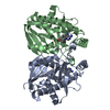
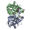
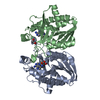
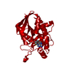
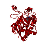
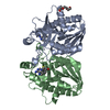
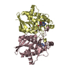
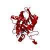
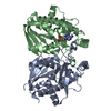
- PDB-1y6q: Cyrstal structure of MTA/AdoHcy nucleosidase complexed with MT-DA... -
+ Open data

- Basic information
| Entry | Database: PDB / ID: 1y6q | ||||||
|---|---|---|---|---|---|---|---|
| Title | Cyrstal structure of MTA/AdoHcy nucleosidase complexed with MT-DADMe-ImmA | ||||||
 Components Components | MTA/SAH nucleosidase | ||||||
 Keywords Keywords | HYDROLASE / mixed alpha/beta dimer | ||||||
| Function / homology |  Function and homology information Function and homology informationpurine deoxyribonucleoside catabolic process / toxic metabolite repair / adenosylhomocysteine nucleosidase / adenosylhomocysteine nucleosidase activity / methylthioadenosine nucleosidase activity / : / : / protein homodimerization activity / identical protein binding / cytosol Similarity search - Function | ||||||
| Biological species |  | ||||||
| Method |  X-RAY DIFFRACTION / FOURIER SYNTHESIS / Resolution: 2.2 Å X-RAY DIFFRACTION / FOURIER SYNTHESIS / Resolution: 2.2 Å | ||||||
 Authors Authors | Lee, J.E. / Singh, V. / Evans, G.B. / Tyler, P.C. / Furneaux, R.H. / Cornell, K.A. / Riscoe, M.K. / Schramm, V.L. / Howell, P.L. | ||||||
 Citation Citation | Journal: J.Biol.Chem. / Year: 2005 Title: Structural rationale for the affinity of pico- and femtomolar transition state analogues of Escherichia coli 5'-methylthioadenosine/S-adenosylhomocysteine nucleosidase. Authors: Lee, J.E. / Singh, V. / Evans, G.B. / Tyler, P.C. / Furneaux, R.H. / Cornell, K.A. / Riscoe, M.K. / Schramm, V.L. / Howell, P.L. #1: Journal: To be PublishedTitle: Femtomolar transition state analogue inhibitors of 5'-methylthioadenosine/S-adenosylhomocysteine nucleosidase from Escherichia coli. Authors: Singh, V. / Evans, G.B. / Lenz, D.H. / Painter, G.F. / Tyler, P.C. / Furneaux, R.H. / Lee, J.E. / Howell, P.L. | ||||||
| History |
|
- Structure visualization
| Structure viewer | Molecule: MolmilJmol/JSmol |
|---|
- Downloads & links
-Download
| PDBx/mmCIF format | 1y6q.cif.gz | 104.4 KB | Display | PDBx/mmCIF format |
|---|---|---|---|---|
| PDB format | pdb1y6q.ent.gz | 79.7 KB | Display | PDB format |
| PDBx/mmJSON format | 1y6q.json.gz | Tree view | PDBx/mmJSON format | |
| Others |  Other downloads Other downloads |
-Validation report
| Arichive directory | https://data.pdbj.org/pub/pdb/validation_reports/y6/1y6qftp://data.pdbj.org/pub/pdb/validation_reports/y6/1y6q | HTTPS FTP |
|---|
-Related structure data
| Related structure data | 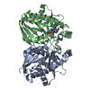 1y6rC  1nc1S S: Starting model for refinement C: citing same article ( |
|---|---|
| Similar structure data |

-Links
 PDBj
PDBj- Assembly
| Deposited unit | 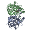
| ||||||||
|---|---|---|---|---|---|---|---|---|---|
| 1 |
| ||||||||
| Unit cell |
|
-Components
| #1: Protein | Mass: 25488.123 Da / Num. of mol.: 2 Source method: isolated from a genetically manipulated source Source: (gene. exp.) References: UniProt: P24247, UniProt: P0AF12*PLUS, methylthioadenosine nucleosidase, adenosylhomocysteine nucleosidase #2: Chemical |   Mass: 35.453 Da / Num. of mol.: 2 / Source method: obtained synthetically / Formula: Cl Mass: 35.453 Da / Num. of mol.: 2 / Source method: obtained synthetically / Formula: Cl#3: Chemical | 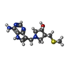  Mass: 293.388 Da / Num. of mol.: 2 / Source method: obtained synthetically / Formula: C13H19N5OS Mass: 293.388 Da / Num. of mol.: 2 / Source method: obtained synthetically / Formula: C13H19N5OS#4: Water | ChemComp-HOH / |  Mass: 18.015 Da / Num. of mol.: 219 / Source method: isolated from a natural source / Formula: H2O Mass: 18.015 Da / Num. of mol.: 219 / Source method: isolated from a natural source / Formula: H2O |
|---|
-Experimental details
-Experiment
| Experiment | Method: X-RAY DIFFRACTION / Number of used crystals: 1 |
|---|
- Sample preparation
| Crystal | Density Matthews: 2.3 Å3/Da / Density % sol: 45.8 % |
|---|---|
| Crystal grow | Temperature: 293 K / Method: vapor diffusion, hanging drop / pH: 4.6 Details: sodium chloride, sodium acetate, pH 4.6, VAPOR DIFFUSION, HANGING DROP, temperature 293K |
-Data collection
| Diffraction | Mean temperature: 100 K |
|---|---|
| Diffraction source | Source: ROTATING ANODE / Type: RIGAKU RU300 / Wavelength: 1.5418 Å |
| Detector | Type: RIGAKU RAXIS IV / Detector: IMAGE PLATE / Date: Nov 4, 2002 / Details: mirrors |
| Radiation | Monochromator: Yale Mirrors / Protocol: SINGLE WAVELENGTH / Monochromatic (M) / Laue (L): M / Scattering type: x-ray |
| Radiation wavelength | Wavelength: 1.5418 Å / Relative weight: 1 |
| Reflection | Resolution: 2.2→28.22 Å / Num. obs: 24071 / Observed criterion σ(F): 0 / Biso Wilson estimate: 37.6 Å2 / Limit h max: 23 / Limit h min: 0 / Limit k max: 31 / Limit k min: 0 / Limit l max: 58 / Limit l min: 0 / Observed criterion F max: 906752.43 / Observed criterion F min: 9.426 |
| Reflection shell | Resolution: 2.2→2.28 Å |
- Processing
| Software |
| ||||||||||||||||||||||||||||||||||||||||||||||||||||||||||||||||||||||||||||||||||||||||||
|---|---|---|---|---|---|---|---|---|---|---|---|---|---|---|---|---|---|---|---|---|---|---|---|---|---|---|---|---|---|---|---|---|---|---|---|---|---|---|---|---|---|---|---|---|---|---|---|---|---|---|---|---|---|---|---|---|---|---|---|---|---|---|---|---|---|---|---|---|---|---|---|---|---|---|---|---|---|---|---|---|---|---|---|---|---|---|---|---|---|---|---|
| Refinement | Method to determine structure: FOURIER SYNTHESIS Starting model: PDB ENTRY 1NC1 Resolution: 2.2→28.22 Å / Rfactor Rfree error: 0.007 / Occupancy max: 1 / Occupancy min: 0 / Cross valid method: THROUGHOUT / σ(F): 0 / σ(I): 0 / Stereochemistry target values: Engh & Huber
| ||||||||||||||||||||||||||||||||||||||||||||||||||||||||||||||||||||||||||||||||||||||||||
| Solvent computation | Solvent model: CNS bulk solvent model used / Bsol: 45.1726 Å2 / ksol: 0.390464 e/Å3 | ||||||||||||||||||||||||||||||||||||||||||||||||||||||||||||||||||||||||||||||||||||||||||
| Displacement parameters | Biso max: 65.04 Å2 / Biso mean: 29.17 Å2 / Biso min: 7.9 Å2
| ||||||||||||||||||||||||||||||||||||||||||||||||||||||||||||||||||||||||||||||||||||||||||
| Refine analyze |
| ||||||||||||||||||||||||||||||||||||||||||||||||||||||||||||||||||||||||||||||||||||||||||
| Refinement step | Cycle: LAST / Resolution: 2.2→28.22 Å
| ||||||||||||||||||||||||||||||||||||||||||||||||||||||||||||||||||||||||||||||||||||||||||
| Refine LS restraints |
| ||||||||||||||||||||||||||||||||||||||||||||||||||||||||||||||||||||||||||||||||||||||||||
| LS refinement shell | Refine-ID: X-RAY DIFFRACTION
|
