 Movie
Movie Controller

[English] 日本語
 Yorodumi
- PDB-3df9: Crystal structure of E. coli MTA/SAH nucleosidase in complex with... -
+ Open data

- Basic information
| Entry | Database: PDB / ID: 3df9 | ||||||
|---|---|---|---|---|---|---|---|
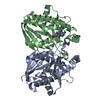
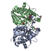
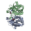
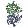
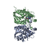
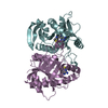
| Title | Crystal structure of E. coli MTA/SAH nucleosidase in complex with BnT-DADMeImmA | ||||||
 Components Components | MTA/SAH nucleosidase | ||||||
 Keywords Keywords | HYDROLASE / mixed alpha/beta dimer | ||||||
| Function / homology |  Function and homology information Function and homology informationpurine deoxyribonucleoside catabolic process / adenosylhomocysteine nucleosidase / adenosylhomocysteine nucleosidase activity / methylthioadenosine nucleosidase activity / : / : / cytosol Similarity search - Function | ||||||
| Biological species |  | ||||||
| Method |  X-RAY DIFFRACTION / MOLECULAR REPLACEMENT / Resolution: 1.95 Å X-RAY DIFFRACTION / MOLECULAR REPLACEMENT / Resolution: 1.95 Å | ||||||
 Authors Authors | Siu, K.K.W. / Howell, P.L. | ||||||
 Citation Citation | Journal: Acta Crystallogr.,Sect.F / Year: 2008 Title: Structure of Staphylococcus aureus 5'-methylthioadenosine/S-adenosylhomocysteine nucleosidase Authors: Siu, K.K. / Lee, J.E. / Smith, G.D. / Horvatin-Mrakovcic, C. / Howell, P.L. | ||||||
| History |
|
- Structure visualization
| Structure viewer | Molecule: MolmilJmol/JSmol |
|---|
- Downloads & links
-Download
| PDBx/mmCIF format | 3df9.cif.gz | 107.1 KB | Display | PDBx/mmCIF format |
|---|---|---|---|---|
| PDB format | pdb3df9.ent.gz | 82 KB | Display | PDB format |
| PDBx/mmJSON format | 3df9.json.gz | Tree view | PDBx/mmJSON format | |
| Others |  Other downloads Other downloads |
-Validation report
| Arichive directory | https://data.pdbj.org/pub/pdb/validation_reports/df/3df9ftp://data.pdbj.org/pub/pdb/validation_reports/df/3df9 | HTTPS FTP |
|---|
-Related structure data
| Related structure data | 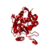 3bl6C 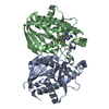 1y6qS S: Starting model for refinement C: citing same article ( |
|---|---|
| Similar structure data |


-Links
 PDBj
PDBj- Assembly
| Deposited unit | 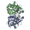
| ||||||||
|---|---|---|---|---|---|---|---|---|---|
| 1 |
| ||||||||
| Unit cell |
|
-Components
| #1: Protein | Mass: 25488.123 Da / Num. of mol.: 2 Source method: isolated from a genetically manipulated source Source: (gene. exp.) References: UniProt: P0AF14, adenosylhomocysteine nucleosidase #2: Chemical |   Mass: 369.484 Da / Num. of mol.: 2 / Source method: obtained synthetically / Formula: C19H23N5OS Mass: 369.484 Da / Num. of mol.: 2 / Source method: obtained synthetically / Formula: C19H23N5OS#3: Water | ChemComp-HOH / |  Mass: 18.015 Da / Num. of mol.: 299 / Source method: isolated from a natural source / Formula: H2O Mass: 18.015 Da / Num. of mol.: 299 / Source method: isolated from a natural source / Formula: H2O |
|---|
-Experimental details
-Experiment
| Experiment | Method: X-RAY DIFFRACTION / Number of used crystals: 1 |
|---|
- Sample preparation
| Crystal | Density Matthews: 2.26 Å3/Da / Density % sol: 45.51 % |
|---|---|
| Crystal grow | Temperature: 298 K / Method: vapor diffusion, hanging drop / pH: 4.5 Details: 35 % (v/v) 2-propanol, 50 mM sodium acetate, pH 4.5, VAPOR DIFFUSION, HANGING DROP, temperature 298K |
-Data collection
| Diffraction | Mean temperature: 100 K |
|---|---|
| Diffraction source | Source: ROTATING ANODE / Type: RIGAKU RU300 / Wavelength: 1.5418 Å |
| Detector | Type: RIGAKU RAXIS IV / Detector: IMAGE PLATE / Date: Jul 20, 2005 |
| Radiation | Monochromator: Confocal multilayer / Protocol: SINGLE WAVELENGTH / Monochromatic (M) / Laue (L): M / Scattering type: x-ray |
| Radiation wavelength | Wavelength: 1.5418 Å / Relative weight: 1 |
| Reflection | Resolution: 1.95→63.93 Å / Num. all: 34475 / Num. obs: 33372 / % possible obs: 96.8 % / Observed criterion σ(F): 5 / Observed criterion σ(I): 5 / Redundancy: 15.9 % / Rmerge(I) obs: 0.078 / Net I/σ(I): 24.6 |
| Reflection shell | Resolution: 1.95→2.02 Å / Redundancy: 15.9 % / Rmerge(I) obs: 0.3 / Mean I/σ(I) obs: 9.8 / % possible all: 95.3 |
- Processing
| Software |
| |||||||||||||||||||||||||||||||||||||||||||||||||||||||||||||||||
|---|---|---|---|---|---|---|---|---|---|---|---|---|---|---|---|---|---|---|---|---|---|---|---|---|---|---|---|---|---|---|---|---|---|---|---|---|---|---|---|---|---|---|---|---|---|---|---|---|---|---|---|---|---|---|---|---|---|---|---|---|---|---|---|---|---|---|
| Refinement | Method to determine structure: MOLECULAR REPLACEMENT Starting model: 1Y6Q Resolution: 1.95→63.89 Å / Cor.coef. Fo:Fc: 0.95 / Cor.coef. Fo:Fc free: 0.937 / SU B: 3.384 / SU ML: 0.098 / Cross valid method: THROUGHOUT / ESU R: 0.171 / ESU R Free: 0.145 / Stereochemistry target values: MAXIMUM LIKELIHOOD / Details: HYDROGENS HAVE BEEN ADDED IN THE RIDING POSITIONS
| |||||||||||||||||||||||||||||||||||||||||||||||||||||||||||||||||
| Solvent computation | Ion probe radii: 0.8 Å / Shrinkage radii: 0.8 Å / VDW probe radii: 1.4 Å / Solvent model: MASK | |||||||||||||||||||||||||||||||||||||||||||||||||||||||||||||||||
| Displacement parameters | Biso mean: 17.654 Å2
| |||||||||||||||||||||||||||||||||||||||||||||||||||||||||||||||||
| Refinement step | Cycle: LAST / Resolution: 1.95→63.89 Å
| |||||||||||||||||||||||||||||||||||||||||||||||||||||||||||||||||
| Refine LS restraints |
| |||||||||||||||||||||||||||||||||||||||||||||||||||||||||||||||||
| LS refinement shell | Resolution: 1.95→2.001 Å / Total num. of bins used: 20
|
