- PDB-4yme: Crystal structure of a sensory box/GGDEF family protein (CC_0091)... -
+
Open data
ID or keywords:
Loading...
-
Basic information
Entry
Database: PDB / ID: 4yme
Title
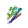
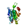
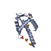
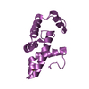
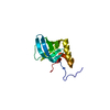
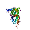
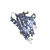
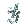
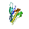
Crystal structure of a sensory box/GGDEF family protein (CC_0091) from Caulobacter crescentus CB15 at 1.40 A resolution (PSI Community Target, Shapiro)
Components
sensory box/GGDEF family protein
Keywords
LYASE / GGDEF domain / PF00990 family / ferredoxin-like fold / Structural Genomics / Joint Center for Structural Genomics / JCSG / Protein Structure Initiative / PSI-BIOLOGY
Mass: 18.015 Da / Num. of mol.: 186 / Source method: isolated from a natural source / Formula: H2O
Has protein modification
Y
Sequence details
THE CONSTRUCT (372-528) WAS EXPRESSED WITH A PURIFICATION TAG MGSDKIHHHHHHENLYFQG. THE TAG WAS ...THE CONSTRUCT (372-528) WAS EXPRESSED WITH A PURIFICATION TAG MGSDKIHHHHHHENLYFQG. THE TAG WAS REMOVED WITH TEV PROTEASE LEAVING ONLY A GLYCINE (0) FOLLOWED BY THE TARGET SEQUENCE.
-
Experimental details
-
Experiment
Experiment
Method: X-RAY DIFFRACTION / Number of used crystals: 1
-
Sample preparation
Crystal
Density Matthews: 2.11 Å3/Da / Density % sol: 41.67 %
Crystal grow
Temperature: 277 K / Method: vapor diffusion, sitting drop Details: 0.2M magnesium formate, 20% polyethylene glycol 3350
Monochromator: double crystal Si(111) / Protocol: SINGLE WAVELENGTH / Monochromatic (M) / Laue (L): M / Scattering type: x-ray
Radiation wavelength
Wavelength: 0.97952 Å / Relative weight: 1
Reflection
Resolution: 1.4→23.679 Å / Num. obs: 30788 / % possible obs: 98.3 % / Observed criterion σ(I): -3 / Redundancy: 6.17 % / Biso Wilson estimate: 19.725 Å2 / Rmerge F obs: 0.999 / Rmerge(I) obs: 0.03 / Rrim(I) all: 0.038 / Net I/σ(I): 15.62 / Num. measured all: 145154
Reflection shell
Resolution (Å)
Highest resolution (Å)
Rmerge F obs
Rmerge(I) obs
Mean I/σ(I) obs
Num. measured obs
Num. possible
Num. unique obs
Rrim(I) all
Diffraction-ID
% possible all
1.4-1.45
0.563
0.946
1.4
14061
5849
5650
1.187
1
96.6
1.45-1.51
0.794
0.568
2.3
14933
6049
6007
0.716
99.3
1.51-1.58
0.921
0.338
3.7
14614
5883
5858
0.425
99.6
1.58-1.66
0.959
0.223
5.3
14096
5635
5612
0.28
99.6
1.66-1.76
0.978
0.155
7.4
14190
5660
5625
0.194
99.4
1.76-1.9
0.993
0.087
11.6
15090
6028
5971
0.11
99.1
1.9-2.09
0.997
0.048
18.8
14503
5819
5729
0.06
98.5
2.09-2.39
0.998
0.03
27.9
14742
5863
5785
0.039
98.7
2.39-3.01
0.999
0.022
35.4
14676
5858
5743
0.028
98
3.01
0.999
0.018
43.5
14249
5931
5581
0.023
94.1
-
Phasing
Phasing
Method: SAD
-
Processing
Software
Name
Version
Classification
PDB_EXTRACT
3.1
dataextraction
SHELX
phasing
SHARP
phasing
XDS
November3, 2014BUILT=20141118
datascaling
REFMAC
5.8.0103
refinement
XSCALE
datascaling
SHELXD
phasing
Refinement
Method to determine structure: SAD / Resolution: 1.4→23.679 Å / Cor.coef. Fo:Fc: 0.969 / Cor.coef. Fo:Fc free: 0.956 / Occupancy max: 1 / Occupancy min: 0.23 / SU B: 2.939 / SU ML: 0.054 / Cross valid method: THROUGHOUT / σ(F): 0 / ESU R: 0.064 / ESU R Free: 0.064 Stereochemistry target values: MAXIMUM LIKELIHOOD WITH PHASES Details: 1. HYDROGENS HAVE BEEN ADDED IN THE RIDING POSITIONS. 2. ATOM RECORDS CONTAIN SUM OF TLS AND RESIDUAL B FACTORS. 3. ANISOU RECORDS CONTAIN SUM OF TLS AND RESIDUAL U FACTORS. 4. A MET- ...Details: 1. HYDROGENS HAVE BEEN ADDED IN THE RIDING POSITIONS. 2. ATOM RECORDS CONTAIN SUM OF TLS AND RESIDUAL B FACTORS. 3. ANISOU RECORDS CONTAIN SUM OF TLS AND RESIDUAL U FACTORS. 4. A MET-INHIBITION PROTOCOL WAS USED FOR SELENOMETHIONINE INCORPORATION DURING PROTEIN EXPRESSION. THE OCCUPANCY OF THE SE ATOMS IN THE MSE RESIDUES WAS REDUCED TO 0.75 FOR THE REDUCED SCATTERING POWER DUE TO PARTIAL S-MET INCORPORATION. 5. THE SAD PHASES WERE USED AS RESTRAINTS DURING REFINEMENT
Rfactor
Num. reflection
% reflection
Selection details
Rfree
0.2048
1546
5 %
RANDOM
Rwork
0.1827
29187
-
-
obs
0.1839
30733
99 %
-
Solvent computation
Ion probe radii: 0.8 Å / Shrinkage radii: 0.8 Å / VDW probe radii: 1.2 Å / Solvent model: MASK
In the structure databanks used in Yorodumi, some data are registered as the other names, "COVID-19 virus" and "2019-nCoV". Here are the details of the virus and the list of structure data.
Jan 31, 2019. EMDB accession codes are about to change! (news from PDBe EMDB page)
EMDB accession codes are about to change! (news from PDBe EMDB page)
The allocation of 4 digits for EMDB accession codes will soon come to an end. Whilst these codes will remain in use, new EMDB accession codes will include an additional digit and will expand incrementally as the available range of codes is exhausted. The current 4-digit format prefixed with “EMD-” (i.e. EMD-XXXX) will advance to a 5-digit format (i.e. EMD-XXXXX), and so on. It is currently estimated that the 4-digit codes will be depleted around Spring 2019, at which point the 5-digit format will come into force.
The EM Navigator/Yorodumi systems omit the EMD- prefix.
Related info.:Q: What is EMD? / ID/Accession-code notation in Yorodumi/EM Navigator
Yorodumi is a browser for structure data from EMDB, PDB, SASBDB, etc.
This page is also the successor to EM Navigator detail page, and also detail information page/front-end page for Omokage search.
The word "yorodu" (or yorozu) is an old Japanese word meaning "ten thousand". "mi" (miru) is to see.
Related info.:EMDB / PDB / SASBDB / Comparison of 3 databanks / Yorodumi Search / Aug 31, 2016. New EM Navigator & Yorodumi / Yorodumi Papers / Jmol/JSmol / Function and homology information / Changes in new EM Navigator and Yorodumi
 Movie
Movie Controller

 Yorodumi
 Open data
 Basic information
 Components
 Keywords
 Function and homology information
 Caulobacter crescentus (bacteria)
 X-RAY DIFFRACTION /
  Authors
 Citation
 Structure visualization
 Downloads & links
 Other downloads
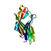
 PDBj
PDBj


 Assembly
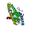
 Mass: 18.015 Da / Num. of mol.: 186 / Source method: isolated from a natural source / Formula: H2O
 Sample preparation
 / Beamline: BL14-1 / Wavelength: 0.97952 Å
 Processing