 Movie
Movie Controller


+ Open data

- Basic information
| Entry | Database: PDB / ID: 5y7z | ||||||
|---|---|---|---|---|---|---|---|
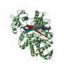
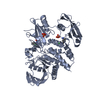
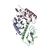
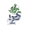
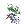
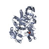
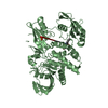
| Title | Complex structure of cyclin G-associated kinase with gefitinib | ||||||
 Components Components |
| ||||||
 Keywords Keywords | TRANSFERASE/IMMUNE SYSTEM / Kinase / complex / TRANSFERASE-IMMUNE SYSTEM complex | ||||||
| Function / homology |  Function and homology information Function and homology informationregulation of clathrin coat assembly / synaptic vesicle uncoating / Golgi to lysosome transport / protein localization to Golgi apparatus / clathrin coat disassembly / clathrin coat assembly / clathrin-dependent endocytosis / endoplasmic reticulum organization / clathrin-coated vesicle / clathrin binding ...regulation of clathrin coat assembly / synaptic vesicle uncoating / Golgi to lysosome transport / protein localization to Golgi apparatus / clathrin coat disassembly / clathrin coat assembly / clathrin-dependent endocytosis / endoplasmic reticulum organization / clathrin-coated vesicle / clathrin binding / Golgi organization / Golgi Associated Vesicle Biogenesis / intracellular transport / protein folding chaperone / receptor-mediated endocytosis / cyclin binding / protein localization to plasma membrane / negative regulation of neuron projection development / Clathrin-mediated endocytosis / presynapse / protein folding / vesicle / non-specific serine/threonine protein kinase / protein serine kinase activity / focal adhesion / protein serine/threonine kinase activity / perinuclear region of cytoplasm / Golgi apparatus / ATP binding / membrane / cytosol / cytoplasm Similarity search - Function | ||||||
| Biological species |  Homo sapiens (human) Homo sapiens (human) | ||||||
| Method |  X-RAY DIFFRACTION / SYNCHROTRON / MOLECULAR REPLACEMENT / Resolution: 2.804 Å X-RAY DIFFRACTION / SYNCHROTRON / MOLECULAR REPLACEMENT / Resolution: 2.804 Å | ||||||
 Authors Authors | Ohbayashi, N. / Murayama, K. / Kato-Murayama, M. / Shirouzu, M. | ||||||
 Citation Citation | Journal: ChemistryOpen / Year: 2018 Title: Structural Basis for the Inhibition of Cyclin G-Associated Kinase by Gefitinib. Authors: Ohbayashi, N. / Murayama, K. / Kato-Murayama, M. / Kukimoto-Niino, M. / Uejima, T. / Matsuda, T. / Ohsawa, N. / Yokoyoma, S. / Nojima, H. / Shirouzu, M. | ||||||
| History |
|
- Structure visualization
| Structure viewer | Molecule: MolmilJmol/JSmol |
|---|
- Downloads & links
-Download
| PDBx/mmCIF format | 5y7z.cif.gz | 184.3 KB | Display | PDBx/mmCIF format |
|---|---|---|---|---|
| PDB format | pdb5y7z.ent.gz | 143.9 KB | Display | PDB format |
| PDBx/mmJSON format | 5y7z.json.gz | Tree view | PDBx/mmJSON format | |
| Others |  Other downloads Other downloads |
-Validation report
| Arichive directory | https://data.pdbj.org/pub/pdb/validation_reports/y7/5y7zftp://data.pdbj.org/pub/pdb/validation_reports/y7/5y7z | HTTPS FTP |
|---|
-Related structure data
| Related structure data | 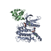 5y80C 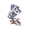 4c57S S: Starting model for refinement C: citing same article ( |
|---|---|
| Similar structure data |


-Links
 PDBj
PDBj


- Assembly
| Deposited unit | 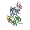
| ||||||||
|---|---|---|---|---|---|---|---|---|---|
| 1 | 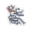
| ||||||||
| 2 | 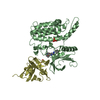
| ||||||||
| Unit cell |
|
-Components
| #1: Protein | Mass: 35657.766 Da / Num. of mol.: 2 / Fragment: UNP RESIDUES 25-335 Source method: isolated from a genetically manipulated source Source: (gene. exp.) Homo sapiens (human) / Gene: GAK / Details (production host): cell free protein synthesis / Production host:  References: UniProt: O14976, non-specific serine/threonine protein kinase #2: Antibody | Mass: 16189.729 Da / Num. of mol.: 2 Source method: isolated from a genetically manipulated source Source: (gene. exp.) #3: Chemical |   Mass: 446.902 Da / Num. of mol.: 2 / Source method: obtained synthetically / Formula: C22H24ClFN4O3 / Feature type: SUBJECT OF INVESTIGATION Mass: 446.902 Da / Num. of mol.: 2 / Source method: obtained synthetically / Formula: C22H24ClFN4O3 / Feature type: SUBJECT OF INVESTIGATION#4: Chemical | ChemComp-SO4 /   Mass: 96.063 Da / Num. of mol.: 5 / Source method: obtained synthetically / Formula: SO4 Mass: 96.063 Da / Num. of mol.: 5 / Source method: obtained synthetically / Formula: SO4#5: Water | ChemComp-HOH / |  Mass: 18.015 Da / Num. of mol.: 9 / Source method: isolated from a natural source / Formula: H2O Mass: 18.015 Da / Num. of mol.: 9 / Source method: isolated from a natural source / Formula: H2OHas protein modification | Y | |
|---|
-Experimental details
-Experiment
| Experiment | Method: X-RAY DIFFRACTION / Number of used crystals: 1 |
|---|
- Sample preparation
| Crystal | Density Matthews: 2.35 Å3/Da / Density % sol: 47.57 % |
|---|---|
| Crystal grow | Temperature: 293 K / Method: vapor diffusion, sitting drop / pH: 6.5 Details: 0.17 M ammonium sulfate, 0.085 M sodium cacodylate trihydrate (pH 6.5), 25.5% PEG 8000, 15% glycerol |
-Data collection
| Diffraction | Mean temperature: 100 K |
|---|---|
| Diffraction source | Source: SYNCHROTRON / Site: SPring-8  / Beamline: BL32XU / Wavelength: 1 Å / Beamline: BL32XU / Wavelength: 1 Å |
| Detector | Type: RAYONIX MX-225 / Detector: CCD / Date: Jul 22, 2014 |
| Radiation | Protocol: SINGLE WAVELENGTH / Monochromatic (M) / Laue (L): M / Scattering type: x-ray |
| Radiation wavelength | Wavelength: 1 Å / Relative weight: 1 |
| Reflection | Resolution: 2.8→50 Å / Num. obs: 23845 / % possible obs: 97 % / Redundancy: 3.3 % / Net I/σ(I): 5.3 |
| Reflection shell | Resolution: 2.8→2.87 Å / Redundancy: 3 % / Mean I/σ(I) obs: 1.46 / Num. unique obs: 1587 / % possible all: 98.6 |
- Processing
| Software |
| ||||||||||||||||||||||||||||||||||||||||||||||||||||||||||||||||||||||
|---|---|---|---|---|---|---|---|---|---|---|---|---|---|---|---|---|---|---|---|---|---|---|---|---|---|---|---|---|---|---|---|---|---|---|---|---|---|---|---|---|---|---|---|---|---|---|---|---|---|---|---|---|---|---|---|---|---|---|---|---|---|---|---|---|---|---|---|---|---|---|---|
| Refinement | Method to determine structure: MOLECULAR REPLACEMENT Starting model: 4C57 Resolution: 2.804→45.251 Å / SU ML: 0.43 / Cross valid method: FREE R-VALUE / σ(F): 1.34 / Phase error: 34.08
| ||||||||||||||||||||||||||||||||||||||||||||||||||||||||||||||||||||||
| Solvent computation | Shrinkage radii: 0.9 Å / VDW probe radii: 1.11 Å | ||||||||||||||||||||||||||||||||||||||||||||||||||||||||||||||||||||||
| Refinement step | Cycle: LAST / Resolution: 2.804→45.251 Å
| ||||||||||||||||||||||||||||||||||||||||||||||||||||||||||||||||||||||
| Refine LS restraints |
| ||||||||||||||||||||||||||||||||||||||||||||||||||||||||||||||||||||||
| LS refinement shell |
|
