 Movie
Movie Controller

[English] 日本語
 Yorodumi
- PDB-5hzy: Crystal structure of the legionella pneumophila effector protein ... -
+ Open data

- Basic information
| Entry | Database: PDB / ID: 5hzy | ||||||
|---|---|---|---|---|---|---|---|
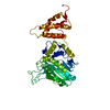
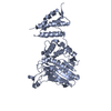
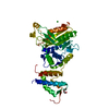
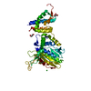
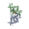
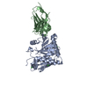
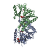
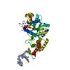
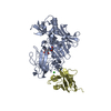
| Title | Crystal structure of the legionella pneumophila effector protein RavZ - P6322 | ||||||
 Components Components | Uncharacterized protein RavZ | ||||||
 Keywords Keywords | HYDROLASE / Autophagy / Atg8 / deconjugating enzyme / protease / Atg4B | ||||||
| Function / homology |  Function and homology information Function and homology informationhost intracellular membrane-bounded organelle / protein delipidation / symbiont-mediated suppression of host autophagy / phosphatidylinositol-3-phosphate binding / cysteine-type peptidase activity / host cell cytoplasmic vesicle membrane / Hydrolases; Acting on peptide bonds (peptidases); Cysteine endopeptidases / proteolysis / extracellular region Similarity search - Function | ||||||
| Biological species |  Legionella pneumophila subsp. pneumophila strain Philadelphia 1 (bacteria) Legionella pneumophila subsp. pneumophila strain Philadelphia 1 (bacteria) | ||||||
| Method |  X-RAY DIFFRACTION / SYNCHROTRON / MOLECULAR REPLACEMENT / Resolution: 2.548 Å X-RAY DIFFRACTION / SYNCHROTRON / MOLECULAR REPLACEMENT / Resolution: 2.548 Å | ||||||
 Authors Authors | Kwon, D.H. / Kim, L. / Kim, B.-W. / Hong, S.B. / Song, H.K. | ||||||
 Citation Citation | Journal: Autophagy / Year: 2017 Title: The 1:2 complex between RavZ and LC3 reveals a mechanism for deconjugation of LC3 on the phagophore membrane Authors: Kwon, D.H. / Kim, S. / Jung, Y.O. / Roh, K.H. / Kim, L. / Kim, B.-W. / Hong, S.B. / Lee, I.Y. / Song, J.H. / Lee, W.C. / Choi, E.J. / Hwang, K.Y. / Song, H.K. | ||||||
| History |
|
- Structure visualization
| Structure viewer | Molecule: MolmilJmol/JSmol |
|---|
- Downloads & links
-Download
| PDBx/mmCIF format | 5hzy.cif.gz | 168.2 KB | Display | PDBx/mmCIF format |
|---|---|---|---|---|
| PDB format | pdb5hzy.ent.gz | 133 KB | Display | PDB format |
| PDBx/mmJSON format | 5hzy.json.gz | Tree view | PDBx/mmJSON format | |
| Others |  Other downloads Other downloads |
-Validation report
| Arichive directory | https://data.pdbj.org/pub/pdb/validation_reports/hz/5hzyftp://data.pdbj.org/pub/pdb/validation_reports/hz/5hzy | HTTPS FTP |
|---|
-Related structure data


-Links
 PDBj
PDBj
- Assembly
| Deposited unit | 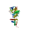
| ||||||||
|---|---|---|---|---|---|---|---|---|---|
| 1 |
| ||||||||
| 2 | 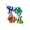
| ||||||||
| Unit cell |
| ||||||||
| Components on special symmetry positions |
|
-Components
| #1: Protein | Mass: 52805.449 Da / Num. of mol.: 1 / Fragment: UNP residues 49-502 / Mutation: C258S Source method: isolated from a genetically manipulated source Source: (gene. exp.) Legionella pneumophila subsp. pneumophila strain Philadelphia 1 (bacteria)Strain: Philadelphia 1 / Gene: lpg1683 / Production host: |
|---|---|
| #2: Water | ChemComp-HOH /  Mass: 18.015 Da / Num. of mol.: 65 / Source method: isolated from a natural source / Formula: H2O Mass: 18.015 Da / Num. of mol.: 65 / Source method: isolated from a natural source / Formula: H2O |
-Experimental details
-Experiment
| Experiment | Method: X-RAY DIFFRACTION / Number of used crystals: 1 |
|---|
- Sample preparation
| Crystal | Density Matthews: 4.33 Å3/Da / Density % sol: 71.58 % |
|---|---|
| Crystal grow | Temperature: 293 K / Method: vapor diffusion, hanging drop / Details: 2.8M Sodium Acetate trihydrate, 200mM NaCl / PH range: 4.0-5.0 |
-Data collection
| Diffraction | Mean temperature: 100 K |
|---|---|
| Diffraction source | Source: SYNCHROTRON / Site: SPring-8  / Beamline: BL44XU / Wavelength: 0.9 Å / Beamline: BL44XU / Wavelength: 0.9 Å |
| Detector | Type: MAR scanner 300 mm plate / Detector: IMAGE PLATE / Date: Jul 14, 2015 |
| Radiation | Protocol: SINGLE WAVELENGTH / Monochromatic (M) / Laue (L): M / Scattering type: x-ray |
| Radiation wavelength | Wavelength: 0.9 Å / Relative weight: 1 |
| Reflection | Resolution: 2.548→41.439 Å / Num. obs: 30876 / % possible obs: 100 % / Redundancy: 4.6 % / Net I/σ(I): 47.61 |
- Processing
| Software |
| |||||||||||||||||||||||||||||||||||||||||||||||||||||||||||||||||||||||||||||||||||||||||||||||||||||||||
|---|---|---|---|---|---|---|---|---|---|---|---|---|---|---|---|---|---|---|---|---|---|---|---|---|---|---|---|---|---|---|---|---|---|---|---|---|---|---|---|---|---|---|---|---|---|---|---|---|---|---|---|---|---|---|---|---|---|---|---|---|---|---|---|---|---|---|---|---|---|---|---|---|---|---|---|---|---|---|---|---|---|---|---|---|---|---|---|---|---|---|---|---|---|---|---|---|---|---|---|---|---|---|---|---|---|---|
| Refinement | Method to determine structure: MOLECULAR REPLACEMENT / Resolution: 2.548→41.439 Å / SU ML: 0.27 / Cross valid method: FREE R-VALUE / σ(F): 1.34 / Phase error: 21.08
| |||||||||||||||||||||||||||||||||||||||||||||||||||||||||||||||||||||||||||||||||||||||||||||||||||||||||
| Solvent computation | Shrinkage radii: 0.9 Å / VDW probe radii: 1.11 Å | |||||||||||||||||||||||||||||||||||||||||||||||||||||||||||||||||||||||||||||||||||||||||||||||||||||||||
| Refinement step | Cycle: LAST / Resolution: 2.548→41.439 Å
| |||||||||||||||||||||||||||||||||||||||||||||||||||||||||||||||||||||||||||||||||||||||||||||||||||||||||
| Refine LS restraints |
| |||||||||||||||||||||||||||||||||||||||||||||||||||||||||||||||||||||||||||||||||||||||||||||||||||||||||
| LS refinement shell |
| |||||||||||||||||||||||||||||||||||||||||||||||||||||||||||||||||||||||||||||||||||||||||||||||||||||||||
| Refinement TLS params. | Method: refined / Refine-ID: X-RAY DIFFRACTION
| |||||||||||||||||||||||||||||||||||||||||||||||||||||||||||||||||||||||||||||||||||||||||||||||||||||||||
| Refinement TLS group |
|
