| 登録情報 | データベース: PDB / ID: 6upa
|
|---|
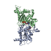
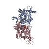
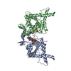
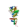
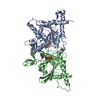
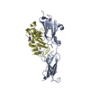
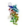
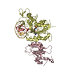
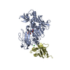
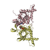
| タイトル | Crystal Structure of GTPase Domain of Human Septin 2/Septin 6 Heterocomplex |
|---|
 要素 要素 | |
|---|
 キーワード キーワード | STRUCTURAL PROTEIN / cytoskeleton protein / septin |
|---|
| 機能・相同性 |  機能・相同性情報 機能・相同性情報
septin collar / septin complex / sperm annulus / photoreceptor connecting cilium / cytoskeleton-dependent cytokinesis / septin ring / smoothened signaling pathway / non-motile cilium / ciliary membrane / cell division site ...septin collar / septin complex / sperm annulus / photoreceptor connecting cilium / cytoskeleton-dependent cytokinesis / septin ring / smoothened signaling pathway / non-motile cilium / ciliary membrane / cell division site / intercellular bridge / cleavage furrow / mitotic cytokinesis / cilium assembly / axoneme / enzyme regulator activity / cytoskeleton organization / Anchoring of the basal body to the plasma membrane / axon terminus / spindle / kinetochore / intracellular protein localization / actin cytoskeleton / microtubule cytoskeleton / synaptic vesicle / midbody / cilium / spermatogenesis / molecular adaptor activity / cell differentiation / cadherin binding / GTPase activity / synapse / GTP binding / cell surface / extracellular exosome / identical protein binding / nucleus / cytoplasm類似検索 - 分子機能 Septin 2 / Septin-type guanine nucleotide-binding (G) domain / Septin / Septin-type guanine nucleotide-binding (G) domain profile. / Septin / P-loop containing nucleoside triphosphate hydrolase類似検索 - ドメイン・相同性 GUANOSINE-5'-DIPHOSPHATE / GUANOSINE-5'-TRIPHOSPHATE / Septin-6 / Septin-2類似検索 - 構成要素 |
|---|
| 生物種 |  Homo sapiens (ヒト) Homo sapiens (ヒト) |
|---|
| 手法 |  X線回折 / シンクロトロン / 分子置換 / 解像度: 2.51 Å X線回折 / シンクロトロン / 分子置換 / 解像度: 2.51 Å |
|---|
 データ登録者 データ登録者 | Rosa, H.V.D. / Brandao-Neto, J. / Martins, C. / Araujo, A.P.U. / Pereira, H.M. / Garratt, R.C. |
|---|
| 資金援助 |  ブラジル, 1件 ブラジル, 1件 | 組織 | 認可番号 | 国 |
|---|
| Sao Paulo Research Foundation (FAPESP) | 2014/15546-1 | ブラジル |
|
|---|
 引用 引用 | ジャーナル: J.Mol.Biol. / 年: 2020
タイトル: Molecular Recognition at Septin Interfaces: The Switches Hold the Key.
著者: Rosa, H.V.D. / Leonardo, D.A. / Brognara, G. / Brandao-Neto, J. / D'Muniz Pereira, H. / Araujo, A.P.U. / Garratt, R.C. |
|---|
| 履歴 | | 登録 | 2019年10月17日 | 登録サイト: RCSB / 処理サイト: RCSB |
|---|
| 改定 1.0 | 2020年9月23日 | Provider: repository / タイプ: Initial release |
|---|
| 改定 1.1 | 2020年9月30日 | Group: Database references / カテゴリ: citation / citation_author
Item: _citation.pdbx_database_id_PubMed / _citation.title ..._citation.pdbx_database_id_PubMed / _citation.title / _citation_author.identifier_ORCID / _citation_author.name |
|---|
| 改定 1.2 | 2020年10月28日 | Group: Database references / カテゴリ: citation
Item: _citation.journal_volume / _citation.page_first / _citation.page_last |
|---|
| 改定 1.3 | 2023年10月11日 | Group: Data collection / Database references / Refinement description
カテゴリ: chem_comp_atom / chem_comp_bond ...chem_comp_atom / chem_comp_bond / database_2 / pdbx_initial_refinement_model
Item: _database_2.pdbx_DOI / _database_2.pdbx_database_accession |
|---|
| 改定 1.4 | 2024年11月20日 | Group: Structure summary
カテゴリ: pdbx_entry_details / pdbx_modification_feature
Item: _pdbx_entry_details.has_protein_modification |
|---|
|
|---|
 ムービー
ムービー コントローラー


 データを開く
 基本情報
基本情報 構造の表示
構造の表示 ダウンロードとリンク
 その他のダウンロード


 PDBj
PDBj 集合体
集合体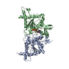


 分子量: 523.180 Da / 分子数: 1 / 由来タイプ: 合成 / 式: C10H16N5O14P3 / タイプ: SUBJECT OF INVESTIGATION / コメント: GTP, エネルギー貯蔵分子*YM
 分子量: 24.305 Da / 分子数: 1 / 由来タイプ: 合成 / 式: Mg / タイプ: SUBJECT OF INVESTIGATION
 タイプ: RNA linking / 分子量: 443.201 Da / 分子数: 1 / 由来タイプ: 合成 / 式: C10H15N5O11P2 / タイプ: SUBJECT OF INVESTIGATION / コメント: GDP, エネルギー貯蔵分子*YM
 試料調製
試料調製 / ビームライン: I24 / 波長: 0.96861 Å
 解析
解析