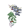
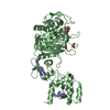
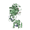
BIOMOLECULE: 1 SEE REMARK 350 FOR THE AUTHOR PROVIDED AND PROGRAM GENERATED ASSEMBLY INFORMATION ... BIOMOLECULE: 1 SEE REMARK 350 FOR THE AUTHOR PROVIDED AND PROGRAM GENERATED ASSEMBLY INFORMATION FOR THE STRUCTURE IN THIS ENTRY. THE REMARK MAY ALSO PROVIDE INFORMATION ON BURIED SURFACE AREA. AUTHORS STATE THAT THE BIOLOGICAL UNIT OF THIS PROTEIN IS UNKNOWN.
Protocol: SINGLE WAVELENGTH / Monochromatic (M) / Laue (L): M / Scattering type: x-ray
Radiation wavelength
Wavelength: 0.9777 Å / Relative weight: 1
Reflection
Resolution: 2.5→20 Å / Num. obs: 22998 / % possible obs: 98.9 % / Redundancy: 7.2 % / Rmerge(I) obs: 0.057 / Χ2: 1.86 / Net I/σ(I): 17.1
Reflection shell
Resolution (Å)
Redundancy (%)
Rmerge(I) obs
Num. unique all
Χ2
Diffraction-ID
% possible all
2.5-2.59
6.5
0.435
2193
0.973
1
95.4
2.59-2.69
7.1
0.372
2251
1.02
1
98.9
2.69-2.81
7.3
0.263
2297
1.103
1
98.9
2.81-2.96
7.3
0.177
2283
1.19
1
99.3
2.96-3.15
7.4
0.121
2303
1.455
1
99.3
3.15-3.39
7.4
0.082
2308
1.73
1
99.5
3.39-3.73
7.2
0.07
2314
2.519
1
99.2
3.73-4.26
7.4
0.053
2316
2.519
1
99.7
4.26-5.35
7.4
0.044
2334
2.696
1
99.7
5.35-22.998
7.1
0.033
2399
3.142
1
99.5
-
Phasing
Phasing
Method: sad
-
Processing
Software
Name
Version
Classification
NB
DENZO
datareduction
SCALEPACK
datascaling
SHELX
phasing
RESOLVE
phasing
REFMAC
refmac_5.2.0019
refinement
PDB_EXTRACT
2
dataextraction
ADSC
Quantum
datacollection
Refinement
Method to determine structure: SAD / Resolution: 2.6→20 Å / Cor.coef. Fo:Fc: 0.934 / Cor.coef. Fo:Fc free: 0.905 / WRfactor Rfree: 0.295 / WRfactor Rwork: 0.231 / SU B: 30.037 / SU ML: 0.29 / TLS residual ADP flag: LIKELY RESIDUAL / Cross valid method: THROUGHOUT / σ(F): 0 / ESU R: 0.491 / ESU R Free: 0.33 / Stereochemistry target values: MAXIMUM LIKELIHOOD Details: 1. Hydrogens have been added in the riding positions. 2. Coot, O, molprobity programs have also been used in the refinement. 3. Atomic B-factors shown are residuals from TLS refinement. 4. ...Details: 1. Hydrogens have been added in the riding positions. 2. Coot, O, molprobity programs have also been used in the refinement. 3. Atomic B-factors shown are residuals from TLS refinement. 4. CAVEAT: residues 84-89 in chain A and 74-90 in chain B could not be reliably assigned in the amino acid sequence and were modeled as alanines.
Rfactor
Num. reflection
% reflection
Selection details
Rfree
0.293
1036
4.9 %
thin shells
Rwork
0.234
-
-
-
all
0.237
-
-
-
obs
-
21100
97.24 %
-
Solvent computation
Ion probe radii: 0.8 Å / Shrinkage radii: 0.8 Å / VDW probe radii: 1.4 Å / Solvent model: MASK
Displacement parameters
Biso mean: 22.672 Å2
Baniso -1
Baniso -2
Baniso -3
1-
-0.87 Å2
0 Å2
-2.21 Å2
2-
-
0.94 Å2
0 Å2
3-
-
-
-3.05 Å2
Refinement step
Cycle: LAST / Resolution: 2.6→20 Å
Protein
Nucleic acid
Ligand
Solvent
Total
Num. atoms
3515
0
60
0
3575
Refine LS restraints
Refine-ID
Type
Dev ideal
Dev ideal target
Number
X-RAY DIFFRACTION
r_bond_refined_d
0.015
0.022
3632
X-RAY DIFFRACTION
r_bond_other_d
0.002
0.02
2413
X-RAY DIFFRACTION
r_angle_refined_deg
1.388
1.982
4921
X-RAY DIFFRACTION
r_angle_other_deg
0.889
3
5882
X-RAY DIFFRACTION
r_dihedral_angle_1_deg
6.062
5
442
X-RAY DIFFRACTION
r_dihedral_angle_2_deg
38.412
23.487
152
X-RAY DIFFRACTION
r_dihedral_angle_3_deg
16.616
15
612
X-RAY DIFFRACTION
r_dihedral_angle_4_deg
17.289
15
24
X-RAY DIFFRACTION
r_chiral_restr
0.073
0.2
580
X-RAY DIFFRACTION
r_gen_planes_refined
0.004
0.02
3925
X-RAY DIFFRACTION
r_gen_planes_other
0.001
0.02
729
X-RAY DIFFRACTION
r_nbd_refined
0.225
0.2
766
X-RAY DIFFRACTION
r_nbd_other
0.196
0.2
2426
X-RAY DIFFRACTION
r_nbtor_refined
0.187
0.2
1757
X-RAY DIFFRACTION
r_nbtor_other
0.087
0.2
1986
X-RAY DIFFRACTION
r_xyhbond_nbd_refined
0.157
0.2
78
X-RAY DIFFRACTION
r_symmetry_vdw_refined
0.188
0.2
10
X-RAY DIFFRACTION
r_symmetry_vdw_other
0.341
0.2
27
X-RAY DIFFRACTION
r_symmetry_hbond_refined
0.179
0.2
1
X-RAY DIFFRACTION
r_mcbond_it
2.435
2
2325
X-RAY DIFFRACTION
r_mcbond_other
0.516
2
908
X-RAY DIFFRACTION
r_mcangle_it
3.431
3
3620
X-RAY DIFFRACTION
r_scbond_it
2.324
2
1495
X-RAY DIFFRACTION
r_scangle_it
3.272
3
1301
LS refinement shell
Refine-ID: X-RAY DIFFRACTION / Total num. of bins used: 20
Resolution (Å)
Rfactor Rfree
Num. reflection Rfree
Rfactor Rwork
Num. reflection Rwork
Num. reflection all
% reflection obs (%)
2.6-2.666
0.447
136
0.346
1366
1558
96.406
2.666-2.738
0
0.308
1479
1508
98.077
2.738-2.816
0.392
119
0.274
1334
1501
96.802
2.816-2.901
0.294
24
0.278
1393
1459
97.121
2.901-2.994
0.337
122
0.266
1253
1409
97.587
2.994-3.096
0
0.241
1330
1365
97.436
3.096-3.21
0.326
91
0.24
1166
1298
96.841
3.21-3.338
0
0.246
1219
1270
95.984
3.338-3.482
0.347
95
0.253
1068
1232
94.399
3.482-3.646
0.344
96
0.276
964
1148
92.334
3.646-3.836
0
0.267
1073
1111
96.58
3.836-4.06
0.316
61
0.218
986
1079
97.034
4.06-4.327
0.224
74
0.2
913
1003
98.405
4.327-4.656
0
0.165
902
906
99.559
4.656-5.073
0.184
74
0.175
807
885
99.548
5.073-5.627
0.243
35
0.214
748
785
99.745
5.627-6.413
0.247
24
0.256
690
715
99.86
6.413-7.66
0.341
53
0.248
560
614
99.837
7.66-10.116
0.377
9
0.208
495
506
99.605
10.116-20
0.244
23
0.254
318
347
98.271
Refinement TLS params.
Method: refined / Refine-ID: X-RAY DIFFRACTION
ID
L11 (°2)
L12 (°2)
L13 (°2)
L22 (°2)
L23 (°2)
L33 (°2)
S11 (Å °)
S12 (Å °)
S13 (Å °)
S21 (Å °)
S22 (Å °)
S23 (Å °)
S31 (Å °)
S32 (Å °)
S33 (Å °)
T11 (Å2)
T12 (Å2)
T13 (Å2)
T22 (Å2)
T23 (Å2)
T33 (Å2)
Origin x (Å)
Origin y (Å)
Origin z (Å)
1
1.1934
0.3281
-2.1672
1.3801
-0.7029
7.7608
0.0373
-0.1126
0.215
0.1794
0.0448
-0.0697
-0.1816
-0.0987
-0.0821
0.2956
0.0341
-0.0321
0.3727
-0.0751
0.1444
59.924
11.799
8.029
2
1.9423
1.721
1.676
3.1958
2.8794
4.5525
0.0432
0.2101
-0.0346
-0.2275
0.0184
-0.2062
-0.1079
0.0148
-0.0615
0.3032
0.0406
0.0407
0.4065
0.035
0.0775
58.671
-1.554
-27.607
Refinement TLS group
ID
Refine-ID
Refine TLS-ID
Auth asym-ID
Auth seq-ID
1
X-RAY DIFFRACTION
1
A
37 - 306
2
X-RAY DIFFRACTION
2
B
36 - 306
+
About Yorodumi
-
News
-
Feb 9, 2022. New format data for meta-information of EMDB entries
New format data for meta-information of EMDB entries
Version 3 of the EMDB header file is now the official format.
The previous official version 1.9 will be removed from the archive.
In the structure databanks used in Yorodumi, some data are registered as the other names, "COVID-19 virus" and "2019-nCoV". Here are the details of the virus and the list of structure data.
Jan 31, 2019. EMDB accession codes are about to change! (news from PDBe EMDB page)
EMDB accession codes are about to change! (news from PDBe EMDB page)
The allocation of 4 digits for EMDB accession codes will soon come to an end. Whilst these codes will remain in use, new EMDB accession codes will include an additional digit and will expand incrementally as the available range of codes is exhausted. The current 4-digit format prefixed with “EMD-” (i.e. EMD-XXXX) will advance to a 5-digit format (i.e. EMD-XXXXX), and so on. It is currently estimated that the 4-digit codes will be depleted around Spring 2019, at which point the 5-digit format will come into force.
The EM Navigator/Yorodumi systems omit the EMD- prefix.
Related info.:Q: What is EMD? / ID/Accession-code notation in Yorodumi/EM Navigator
Yorodumi is a browser for structure data from EMDB, PDB, SASBDB, etc.
This page is also the successor to EM Navigator detail page, and also detail information page/front-end page for Omokage search.
The word "yorodu" (or yorozu) is an old Japanese word meaning "ten thousand". "mi" (miru) is to see.
Related info.:EMDB / PDB / SASBDB / Comparison of 3 databanks / Yorodumi Search / Aug 31, 2016. New EM Navigator & Yorodumi / Yorodumi Papers / Jmol/JSmol / Function and homology information / Changes in new EM Navigator and Yorodumi
 Movie
Movie Controller


 Open data
 Basic information
 Components
 Keywords
 Function and homology information
 Homo sapiens (human)
 X-RAY DIFFRACTION /
  Authors
 Citation
 Structure visualization
 Downloads & links
 Other downloads
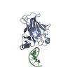


 PDBj
PDBj Assembly
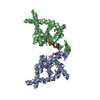


 Type: RNA linking / Mass: 443.201 Da / Num. of mol.: 2 / Source method: obtained synthetically / Formula: C10H15N5O11P2 / Comment: GDP, energy-carrying molecule*YM
 Num. of mol.: 4 / Source method: obtained synthetically
 Sample preparation
 / Beamline: A1 / Wavelength: 0.9777 Å
 Processing