 Movie
Movie Controller

[English] 日本語
 Yorodumi
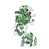
- PDB-3zg8: Crystal Structure of Penicillin Binding Protein 4 from Listeria m... -
+ Open data

- Basic information
| Entry | Database: PDB / ID: 3zg8 | ||||||
|---|---|---|---|---|---|---|---|
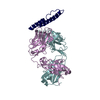
| Title | Crystal Structure of Penicillin Binding Protein 4 from Listeria monocytogenes in the Ampicillin bound form | ||||||
 Components Components |
| ||||||
 Keywords Keywords | PENICILLIN-BINDING PROTEIN | ||||||
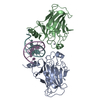
| Function / homology |  Function and homology information Function and homology informationpeptidoglycan glycosyltransferase activity / serine-type D-Ala-D-Ala carboxypeptidase activity / penicillin binding / peptidoglycan biosynthetic process / cell wall organization / regulation of cell shape / outer membrane-bounded periplasmic space / proteolysis Similarity search - Function | ||||||
| Biological species |  LISTERIA MONOCYTOGENES (bacteria) LISTERIA MONOCYTOGENES (bacteria) | ||||||
| Method |  X-RAY DIFFRACTION / SYNCHROTRON / MOLECULAR REPLACEMENT / Resolution: 2.094 Å X-RAY DIFFRACTION / SYNCHROTRON / MOLECULAR REPLACEMENT / Resolution: 2.094 Å | ||||||
 Authors Authors | Jeong, J.H. / Kim, Y.G. | ||||||
 Citation Citation | Journal: Antimicrob.Agents Chemother. / Year: 2013 Title: Crystal Structures of Bifunctional Penicillin-Binding Protein 4 from Listeria Monocytogenes. Authors: Jeong, J. / Kim, Y. / Rojviriya, C. / Ha, S. / Kang, B.S. / Kim, Y. | ||||||
| History |
|
- Structure visualization
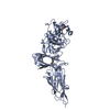
| Structure viewer | Molecule: MolmilJmol/JSmol |
|---|
- Downloads & links
-Download
| PDBx/mmCIF format | 3zg8.cif.gz | 113.6 KB | Display | PDBx/mmCIF format |
|---|---|---|---|---|
| PDB format | pdb3zg8.ent.gz | 85.1 KB | Display | PDB format |
| PDBx/mmJSON format | 3zg8.json.gz | Tree view | PDBx/mmJSON format | |
| Others |  Other downloads Other downloads |
-Validation report
| Arichive directory | https://data.pdbj.org/pub/pdb/validation_reports/zg/3zg8ftp://data.pdbj.org/pub/pdb/validation_reports/zg/3zg8 | HTTPS FTP |
|---|
-Related structure data
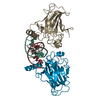
| Related structure data |  3zg7SC  3zgaC S: Starting model for refinement C: citing same article ( |
|---|---|
| Similar structure data |


-Links
 PDBj
PDBj
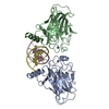
- Assembly
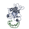
| Deposited unit | 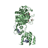
| ||||||||
|---|---|---|---|---|---|---|---|---|---|
| 1 |
| ||||||||
| Unit cell |
|
-Components
| #1: Protein/peptide | Mass: 5049.557 Da / Num. of mol.: 1 / Fragment: RESIDUES 73-119 Source method: isolated from a genetically manipulated source Details: PROTEOLYSIS FROM 120-177 / Source: (gene. exp.) LISTERIA MONOCYTOGENES (bacteria) / Plasmid: PPROEXHTA / Production host: | ||||
|---|---|---|---|---|---|
| #2: Protein | Mass: 58099.547 Da / Num. of mol.: 1 / Fragment: RESIDUES 178-714 Source method: isolated from a genetically manipulated source Source: (gene. exp.) LISTERIA MONOCYTOGENES (bacteria) / Plasmid: PPROEXHTA / Production host: | ||||
| #3: Chemical | ChemComp-AIX / (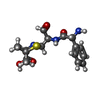  Mass: 351.421 Da / Num. of mol.: 1 / Source method: obtained synthetically / Formula: C16H21N3O4S Mass: 351.421 Da / Num. of mol.: 1 / Source method: obtained synthetically / Formula: C16H21N3O4S | ||||
| #4: Chemical |   Mass: 92.094 Da / Num. of mol.: 3 / Source method: obtained synthetically / Formula: C3H8O3 Mass: 92.094 Da / Num. of mol.: 3 / Source method: obtained synthetically / Formula: C3H8O3#5: Water | ChemComp-HOH / |  Mass: 18.015 Da / Num. of mol.: 121 / Source method: isolated from a natural source / Formula: H2O Mass: 18.015 Da / Num. of mol.: 121 / Source method: isolated from a natural source / Formula: H2OHas protein modification | Y | |
-Experimental details
-Experiment
| Experiment | Method: X-RAY DIFFRACTION / Number of used crystals: 1 |
|---|
- Sample preparation
| Crystal | Density Matthews: 1.91 Å3/Da / Density % sol: 35.75 % / Description: NONE |
|---|---|
| Crystal grow | pH: 7 Details: 20% POLYETHYLENE GLYCOL 3,350 0.1M AMMONIUM TARTRATE (PH 7.0) |
-Data collection
| Diffraction | Mean temperature: 295 K |
|---|---|
| Diffraction source | Source: SYNCHROTRON / Site: PAL/PLS  / Beamline: 4A / Wavelength: 0.9795 / Beamline: 4A / Wavelength: 0.9795 |
| Detector | Type: ADSC QUANTUM 315 / Detector: CCD / Date: Mar 20, 2010 / Details: MIRRORS |
| Radiation | Monochromator: DOUBLE CRYSTAL MONOCHROMATOR / Protocol: SINGLE WAVELENGTH / Monochromatic (M) / Laue (L): M / Scattering type: x-ray |
| Radiation wavelength | Wavelength: 0.9795 Å / Relative weight: 1 |
| Reflection | Resolution: 2.1→30 Å / Num. obs: 35362 / % possible obs: 98.5 % / Observed criterion σ(I): 0 / Redundancy: 5.5 % / Biso Wilson estimate: 32.48 Å2 / Rmerge(I) obs: 0.07 / Net I/σ(I): 27.3 |
| Reflection shell | Resolution: 2.1→2.18 Å / Redundancy: 3.7 % / Rmerge(I) obs: 0.32 / Mean I/σ(I) obs: 4.5 / % possible all: 88.3 |
- Processing
| Software |
| |||||||||||||||||||||||||||||||||||||||||||||||||||||||||||||||||||||||||||||||||||||||||||||||||||||||||
|---|---|---|---|---|---|---|---|---|---|---|---|---|---|---|---|---|---|---|---|---|---|---|---|---|---|---|---|---|---|---|---|---|---|---|---|---|---|---|---|---|---|---|---|---|---|---|---|---|---|---|---|---|---|---|---|---|---|---|---|---|---|---|---|---|---|---|---|---|---|---|---|---|---|---|---|---|---|---|---|---|---|---|---|---|---|---|---|---|---|---|---|---|---|---|---|---|---|---|---|---|---|---|---|---|---|---|
| Refinement | Method to determine structure: MOLECULAR REPLACEMENT Starting model: PDB ENTRY 3ZG7 Resolution: 2.094→29.698 Å / SU ML: 0.27 / σ(F): 1.48 / Phase error: 22.87 / Stereochemistry target values: ML
| |||||||||||||||||||||||||||||||||||||||||||||||||||||||||||||||||||||||||||||||||||||||||||||||||||||||||
| Solvent computation | Shrinkage radii: 0.9 Å / VDW probe radii: 1.11 Å / Solvent model: FLAT BULK SOLVENT MODEL | |||||||||||||||||||||||||||||||||||||||||||||||||||||||||||||||||||||||||||||||||||||||||||||||||||||||||
| Displacement parameters | Biso mean: 25.8 Å2 | |||||||||||||||||||||||||||||||||||||||||||||||||||||||||||||||||||||||||||||||||||||||||||||||||||||||||
| Refinement step | Cycle: LAST / Resolution: 2.094→29.698 Å
| |||||||||||||||||||||||||||||||||||||||||||||||||||||||||||||||||||||||||||||||||||||||||||||||||||||||||
| Refine LS restraints |
| |||||||||||||||||||||||||||||||||||||||||||||||||||||||||||||||||||||||||||||||||||||||||||||||||||||||||
| LS refinement shell |
|
