Protocol: SINGLE WAVELENGTH / Monochromatic (M) / Laue (L): M / Scattering type: x-ray
Radiation wavelength
Wavelength: 0.9686 Å / Relative weight: 1
Reflection
Resolution: 6.6→50 Å / Num. obs: 2508 / % possible obs: 97.7 % / Observed criterion σ(I): 0 / Redundancy: 6 % / Biso Wilson estimate: 121.5 Å2 / Rmerge(I) obs: 0.23 / Net I/σ(I): 7.5
Reflection shell
Resolution: 6.6→6.8 Å / Redundancy: 5 % / Rmerge(I) obs: 0.79 / Mean I/σ(I) obs: 2.3 / % possible all: 97.4
-
Processing
Software
Name
Version
Classification
PHENIX
(PHENIX.REFINE)
refinement
xia2
datareduction
SCALA
datascaling
PHASER
phasing
Refinement
Method to determine structure: MOLECULAR REPLACEMENT / Resolution: 6.601→84.779 Å / SU ML: 0.99 / σ(F): 1.36 / Phase error: 27.17 / Stereochemistry target values: ML Details: THERE IS AN AUTOCATALYTIC CLEAVAGE SITE PRESENT IN RGMB ( CHAINS C AND D), WHICH IS LOCATED BETWEEN RESIDUES ASP168 AND PRO169. IN ADDITION, NEO1 RESIDUES R967 AND R968 IN ( CHAIN A AND B) ...Details: THERE IS AN AUTOCATALYTIC CLEAVAGE SITE PRESENT IN RGMB ( CHAINS C AND D), WHICH IS LOCATED BETWEEN RESIDUES ASP168 AND PRO169. IN ADDITION, NEO1 RESIDUES R967 AND R968 IN ( CHAIN A AND B) WERE BUILT AS ALANINES.
Rfactor
Num. reflection
% reflection
Rfree
0.2799
230
9.2 %
Rwork
0.2537
-
-
obs
0.2561
2488
97.19 %
Solvent computation
Shrinkage radii: 0.9 Å / VDW probe radii: 1.11 Å / Solvent model: FLAT BULK SOLVENT MODEL
Refinement step
Cycle: LAST / Resolution: 6.601→84.779 Å
Protein
Nucleic acid
Ligand
Solvent
Total
Num. atoms
5714
0
0
0
5714
Refine LS restraints
Refine-ID
Type
Dev ideal
Number
X-RAY DIFFRACTION
f_bond_d
0.017
5860
X-RAY DIFFRACTION
f_angle_d
1.934
7972
X-RAY DIFFRACTION
f_dihedral_angle_d
15.753
2126
X-RAY DIFFRACTION
f_chiral_restr
0.132
910
X-RAY DIFFRACTION
f_plane_restr
0.009
1008
LS refinement shell
Resolution (Å)
Rfactor Rfree
Num. reflection Rfree
Rfactor Rwork
Num. reflection Rwork
Refine-ID
% reflection obs (%)
6.6013-8.3158
0.2927
112
0.2839
1116
X-RAY DIFFRACTION
98
8.3158-84.7859
0.2731
118
0.2365
1142
X-RAY DIFFRACTION
97
Refinement TLS params.
Method: refined / Refine-ID: X-RAY DIFFRACTION
ID
L11 (°2)
L12 (°2)
L13 (°2)
L22 (°2)
L23 (°2)
L33 (°2)
S11 (Å °)
S12 (Å °)
S13 (Å °)
S21 (Å °)
S22 (Å °)
S23 (Å °)
S31 (Å °)
S32 (Å °)
S33 (Å °)
T11 (Å2)
T12 (Å2)
T13 (Å2)
T22 (Å2)
T23 (Å2)
T33 (Å2)
Origin x (Å)
Origin y (Å)
Origin z (Å)
1
-2.2328
-0.1047
-3.974
-0.4586
0.9237
2.276
-0.0943
-0.3852
1.0679
-1.2325
0.3497
0.0138
-0.1604
0.6641
0.0127
1.963
0.0278
-0.2478
2.2557
0.2168
2.1172
-32.7187
4.9577
-1.9249
2
3.1885
2.0437
-4.8647
2.0279
2.3967
0.4104
0.2993
0.8024
-0.4103
1.0129
0.1394
0.3895
-0.5137
0.173
0
1.5756
0.2398
-0.4552
1.5913
0.0836
2.0927
-59.2792
-27.2953
10.8912
3
-0.3154
-2.0215
4.7356
-0.3698
-0.8058
-0.56
-0.1838
0.4475
-0.2846
-0.0641
-0.0169
1.1136
0.103
-0.9956
0.0061
2.1725
0.2836
-0.0428
2.6658
0.2728
2.3359
-86.7771
0.2457
-17.3199
4
1.848
0.8582
-1.432
2.643
-1.8267
1.09
-0.3289
-0.0677
0.5234
-0.1219
0.7661
0.1168
-0.5633
0.4283
0
2.6234
0.1082
0.3693
1.5493
0.0001
2.13
-52.1815
19.3293
2.3813
5
0.4262
-0.2097
0.2663
3.9928
1.4585
7.4441
-0.0336
-0.3286
0.3859
0.2892
0.3566
-0.0964
-0.315
0.6012
-0
1.7104
-0.0138
-0.044
1.6487
0.1583
1.8887
-39.3858
-23.6731
20.7424
6
-4.2542
-0.6685
3.4518
6.7632
-3.5219
4.4544
-0.2203
0.2084
-0.2531
0.39
-0.0481
-0.0255
-0.1692
0.4714
0
2.4319
0.2331
0.7155
1.652
-0.1179
1.983
-67.124
7.3282
14.1933
Refinement TLS group
ID
Refine-ID
Refine TLS-ID
Selection details
1
X-RAY DIFFRACTION
1
CHAINAAND (RESID884:983)
2
X-RAY DIFFRACTION
2
CHAINAAND (RESID984:1088)
3
X-RAY DIFFRACTION
3
CHAINBAND (RESID884:983)
4
X-RAY DIFFRACTION
4
CHAINBAND (RESID984:1088)
5
X-RAY DIFFRACTION
5
CHAINCAND (RESID138:321)
6
X-RAY DIFFRACTION
6
CHAINDAND (RESID138:321)
+
About Yorodumi
-
News
-
Feb 9, 2022. New format data for meta-information of EMDB entries
New format data for meta-information of EMDB entries
Version 3 of the EMDB header file is now the official format.
The previous official version 1.9 will be removed from the archive.
In the structure databanks used in Yorodumi, some data are registered as the other names, "COVID-19 virus" and "2019-nCoV". Here are the details of the virus and the list of structure data.
Jan 31, 2019. EMDB accession codes are about to change! (news from PDBe EMDB page)
EMDB accession codes are about to change! (news from PDBe EMDB page)
The allocation of 4 digits for EMDB accession codes will soon come to an end. Whilst these codes will remain in use, new EMDB accession codes will include an additional digit and will expand incrementally as the available range of codes is exhausted. The current 4-digit format prefixed with “EMD-” (i.e. EMD-XXXX) will advance to a 5-digit format (i.e. EMD-XXXXX), and so on. It is currently estimated that the 4-digit codes will be depleted around Spring 2019, at which point the 5-digit format will come into force.
The EM Navigator/Yorodumi systems omit the EMD- prefix.
Related info.:Q: What is EMD? / ID/Accession-code notation in Yorodumi/EM Navigator
Yorodumi is a browser for structure data from EMDB, PDB, SASBDB, etc.
This page is also the successor to EM Navigator detail page, and also detail information page/front-end page for Omokage search.
The word "yorodu" (or yorozu) is an old Japanese word meaning "ten thousand". "mi" (miru) is to see.
Related info.:EMDB / PDB / SASBDB / Comparison of 3 databanks / Yorodumi Search / Aug 31, 2016. New EM Navigator & Yorodumi / Yorodumi Papers / Jmol/JSmol / Function and homology information / Changes in new EM Navigator and Yorodumi
 Movie
Movie Controller


 Open data
 Basic information
 Components
 Keywords
 Function and homology information
 HOMO SAPIENS (human)
 X-RAY DIFFRACTION /
  Authors
 Citation
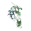
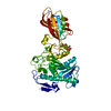
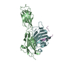
 Structure visualization
 Downloads & links
 Other downloads


 PDBj
PDBj


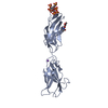
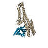
 Assembly
 Sample preparation
 / Beamline: I03 / Wavelength: 0.9686
  Processing