[English] 日本語
Yorodumi
- PDB-4bq6: Crystal structure of the RGMB-NEO1 complex form 1 -

+
Open data


ID or keywords:

Loading...

-
Basic information

Entry
Database: PDB / ID: 4bq6
TitleCrystal structure of the RGMB-NEO1 complex form 1
Components
  • (RGM DOMAIN FAMILY MEMBER B) x 2
  • NEOGENINNEO1
KeywordsCELL ADHESION
Function / homology
Function and homology information


negative regulation of axon regeneration / Netrin-1 signaling / co-receptor binding / BMP receptor binding / regulation of axon regeneration / myoblast fusion / plasma membrane protein complex / positive regulation of BMP signaling pathway / intracellular vesicle / protein secretion ...negative regulation of axon regeneration / Netrin-1 signaling / co-receptor binding / BMP receptor binding / regulation of axon regeneration / myoblast fusion / plasma membrane protein complex / positive regulation of BMP signaling pathway / intracellular vesicle / protein secretion / negative regulation of protein secretion / endoplasmic reticulum-Golgi intermediate compartment / BMP signaling pathway / coreceptor activity / side of membrane / axonal growth cone / axon guidance / neuron migration / cell-cell adhesion / multicellular organismal-level iron ion homeostasis / signaling receptor activity / growth cone / intracellular iron ion homeostasis / cell adhesion / cadherin binding / membrane raft / neuronal cell body / regulation of DNA-templated transcription / positive regulation of DNA-templated transcription / Golgi apparatus / cell surface / signal transduction / nucleoplasm / membrane / identical protein binding / plasma membrane
Similarity search - Function
Repulsive guidance molecule, C-terminal / Repulsive guidance molecule, N-terminal / Repulsive guidance molecule / Repulsive guidance molecule (RGM) C-terminus / Repulsive guidance molecule (RGM) N-terminus / Mog1/PsbP, alpha/beta/alpha sandwich / Protein Transport Mog1p; Chain A / Neogenin, C-terminal / Neogenin C-terminus / Immunoglobulin domain ...Repulsive guidance molecule, C-terminal / Repulsive guidance molecule, N-terminal / Repulsive guidance molecule / Repulsive guidance molecule (RGM) C-terminus / Repulsive guidance molecule (RGM) N-terminus / Mog1/PsbP, alpha/beta/alpha sandwich / Protein Transport Mog1p; Chain A / Neogenin, C-terminal / Neogenin C-terminus / Immunoglobulin domain / Immunoglobulin domain / Immunoglobulin I-set / Immunoglobulin I-set domain / Fibronectin type III domain / Fibronectin type 3 domain / Immunoglobulin subtype 2 / Immunoglobulin C-2 Type / Fibronectin type-III domain profile. / Fibronectin type III / Fibronectin type III superfamily / Immunoglobulin subtype / Immunoglobulin / Ig-like domain profile. / Immunoglobulin-like domain / Immunoglobulin-like domain superfamily / Immunoglobulins / Immunoglobulin-like fold / Immunoglobulin-like / Sandwich / 3-Layer(aba) Sandwich / Mainly Beta / Alpha Beta
Similarity search - Domain/homology
Neogenin / Repulsive guidance molecule B
Similarity search - Component
Biological speciesMUS MUSCULUS (house mouse)
HOMO SAPIENS (human)
MethodX-RAY DIFFRACTION / SYNCHROTRON / MOLECULAR REPLACEMENT / Resolution: 2.3 Å
AuthorsBell, C.H. / Healey, E. / van Erp, S. / Bishop, B. / Tang, C. / Gilbert, R.J.C. / Aricescu, A.R. / Pasterkamp, R.J. / Siebold, C.
CitationJournal: Science / Year: 2013
Title: Structure of the Repulsive Guidance Molecule (Rgm)-Neogenin Signaling Hub
Authors: Bell, C.H. / Healey, E. / Van Erp, S. / Bishop, B. / Tang, C. / Gilbert, R.J.C. / Aricescu, A.R. / Pasterkamp, R.J. / Siebold, C.
History
DepositionMay 30, 2013Deposition site: PDBE / Processing site: PDBE
Revision 1.0Jun 12, 2013Provider: repository / Type: Initial release
Revision 1.1Jun 19, 2013Group: Database references
Revision 1.2Jul 17, 2013Group: Database references
Revision 1.3Apr 3, 2019Group: Data collection / Derived calculations ...Data collection / Derived calculations / Other / Source and taxonomy
Category: entity_src_gen / pdbx_database_proc ...entity_src_gen / pdbx_database_proc / pdbx_database_status / struct_conn
Item: _entity_src_gen.pdbx_host_org_cell_line / _pdbx_database_status.recvd_author_approval / _struct_conn.pdbx_leaving_atom_flag
Revision 1.4Jul 29, 2020Group: Data collection / Derived calculations ...Data collection / Derived calculations / Other / Structure summary
Category: chem_comp / entity ...chem_comp / entity / pdbx_chem_comp_identifier / pdbx_database_status / pdbx_entity_nonpoly / struct_conn / struct_site / struct_site_gen
Item: _chem_comp.name / _chem_comp.type ..._chem_comp.name / _chem_comp.type / _entity.pdbx_description / _pdbx_database_status.status_code_sf / _pdbx_entity_nonpoly.name / _struct_conn.pdbx_role
Description: Carbohydrate remediation / Provider: repository / Type: Remediation

-
Structure visualization

Structure viewerMolecule:
MolmilJmol/JSmol

Downloads & links

-
Assembly

Deposited unit
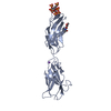
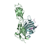
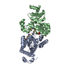
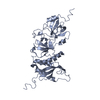
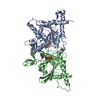
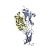
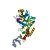
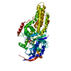
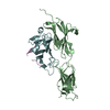
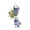
A: NEOGENIN
B: NEOGENIN
C: RGM DOMAIN FAMILY MEMBER B
D: RGM DOMAIN FAMILY MEMBER B
E: RGM DOMAIN FAMILY MEMBER B
F: RGM DOMAIN FAMILY MEMBER B
hetero molecules


Theoretical massNumber of molelcules
Total (without water)141,4148
Polymers140,9726
Non-polymers4422
Water5,927329
1
B: NEOGENIN
E: RGM DOMAIN FAMILY MEMBER B
F: RGM DOMAIN FAMILY MEMBER B
hetero molecules


Theoretical massNumber of molelcules
Total (without water)70,7074
Polymers70,4863
Non-polymers2211
Water181
TypeNameSymmetry operationNumber
identity operation1_555x,y,z1
Buried area4780 Å2
ΔGint-20.6 kcal/mol
Surface area17860 Å2
MethodPISA
2
A: NEOGENIN
C: RGM DOMAIN FAMILY MEMBER B
D: RGM DOMAIN FAMILY MEMBER B
hetero molecules


Theoretical massNumber of molelcules
Total (without water)70,7074
Polymers70,4863
Non-polymers2211
Water543
TypeNameSymmetry operationNumber
identity operation1_555x,y,z1
Buried area4750 Å2
ΔGint-20.5 kcal/mol
Surface area17790 Å2
MethodPISA
Unit cell
Length a, b, c (Å)91.320, 100.390, 103.690
Angle α, β, γ (deg.)90.00, 90.00, 90.00
Int Tables number19
Space group name H-MP212121
Noncrystallographic symmetry (NCS)NCS oper:
IDCodeMatrixVector
1given(-0.9996, 0.01127, -0.0247), (-0.0115, -0.9999, 0.01175), (-0.02456, 0.01203, 0.9996)-23.36, -50.33, -0.1277
2given(-0.9996, -0.003685, -0.02911), (0.003562, -1, 0.004287), (-0.02912, 0.004182, 0.9996)-23.74, -50.6, -0.3116

-
Components

#1: Protein NEOGENIN / NEO1


Mass: 29221.842 Da / Num. of mol.: 2 / Fragment: FN-TYPE III DOMAINS 5 AND 6, RESIDUES 883-1133
Source method: isolated from a genetically manipulated source
Details: N-LINKED GLYCOSYLATION AT N940 / Source: (gene. exp.) MUS MUSCULUS (house mouse) / Plasmid: PHLSEC / Cell line (production host): HEK293T / Production host: HOMO SAPIENS (human) / References: UniProt: P97798
#2: Protein RGM DOMAIN FAMILY MEMBER B / REPULSIVE GUIDANCE MOLECULE B / DRG11-RESPONSIVE AXONAL GUIDANCE AND OUTGROWTH OF NEURITE / DRAGON


Mass: 13318.698 Da / Num. of mol.: 2 / Fragment: ECTODOMAIN, RESIDUES 50-168
Source method: isolated from a genetically manipulated source
Source: (gene. exp.) HOMO SAPIENS (human) / Plasmid: PHLSEC / Cell line (production host): HEK293T / Production host: HOMO SAPIENS (human) / References: UniProt: Q6NW40
#3: Protein RGM DOMAIN FAMILY MEMBER B / REPULSIVE GUIDANCE MOLECULE B / DRG11-RESPONSIVE AXONAL GUIDANCE AND OUTGROWTH OF NEURITE / DRAGON


Mass: 27945.352 Da / Num. of mol.: 2 / Fragment: ECTODOMAIN, RESIDUES 169-410
Source method: isolated from a genetically manipulated source
Source: (gene. exp.) HOMO SAPIENS (human) / Plasmid: PHLSEC / Cell line (production host): HEK293T / Production host: HOMO SAPIENS (human) / References: UniProt: Q6NW40
#4: Sugar ChemComp-NAG / 2-acetamido-2-deoxy-beta-D-glucopyranose / N-Acetylglucosamine


Type: D-saccharide, beta linking / Mass: 221.208 Da / Num. of mol.: 2
Source method: isolated from a genetically manipulated source
Formula: C8H15NO6
IdentifierTypeProgram
DGlcpNAcbCONDENSED IUPAC CARBOHYDRATE SYMBOLGMML 1.0
N-acetyl-b-D-glucopyranosamineCOMMON NAMEGMML 1.0
b-D-GlcpNAcIUPAC CARBOHYDRATE SYMBOLPDB-CARE 1.0
GlcNAcSNFG CARBOHYDRATE SYMBOLGMML 1.0
#5: Water ChemComp-HOH / water / Water


Mass: 18.015 Da / Num. of mol.: 329 / Source method: isolated from a natural source / Formula: H2O
Nonpolymer detailsN-ACETYL-D-GLUCOSAMINE (NAG): N-LINKED GLYCOSYLATION OF NEO1 ASN940
Sequence detailsNEO1 RESIDUES R967 AND R968 WERE BUILT AS ALANINES.

-
Experimental details

-
Experiment

ExperimentMethod: X-RAY DIFFRACTION / Number of used crystals: 1

-
Sample preparation

CrystalDensity % sol: 55 % / Description: NONE
Crystal growpH: 8.5
Details: 0.1 M TRIS-HCL, PH 8.5 0.2 M SODIUM ACETATE 30% PEG4000

-
Data collection

DiffractionMean temperature: 100 K
Diffraction sourceSource: SYNCHROTRON / Site: Diamond / Beamline: I03 / Wavelength: 0.97
RadiationProtocol: SINGLE WAVELENGTH / Monochromatic (M) / Laue (L): M / Scattering type: x-ray
Radiation wavelengthWavelength: 0.97 Å / Relative weight: 1
ReflectionResolution: 2.3→57 Å / Num. obs: 42796 / % possible obs: 99.3 % / Observed criterion σ(I): 0 / Redundancy: 6 % / Biso Wilson estimate: 52.99 Å2 / Rmerge(I) obs: 0.09 / Net I/σ(I): 10.2
Reflection shellResolution: 2.3→2.36 Å / Redundancy: 4 % / Rmerge(I) obs: 0.84 / Mean I/σ(I) obs: 1.3 / % possible all: 95.6

-
Processing

Software
NameVersionClassification
BUSTER2.11.2refinement
xia2data reduction
SCALAdata scaling
PHASERphasing
RefinementMethod to determine structure: MOLECULAR REPLACEMENT / Resolution: 2.3→30 Å / Cor.coef. Fo:Fc: 0.9397 / Cor.coef. Fo:Fc free: 0.9115 / SU R Cruickshank DPI: 0.29 / Cross valid method: THROUGHOUT / σ(F): 0 / SU R Blow DPI: 0.301 / SU Rfree Blow DPI: 0.233 / SU Rfree Cruickshank DPI: 0.232
Details: THERE IS A AUTOCATALYTIC CLEAVAGE SITE PRESENT IN RGMB (CHAINS C/D AND D/E), WHICH IS LOCATED BETWEEN RESIDUES ASP168 AND PRO169. IN ADDITION, NEO1 RESIDUES R967 AND R968 IN (CHAIN A AND B) ...Details: THERE IS A AUTOCATALYTIC CLEAVAGE SITE PRESENT IN RGMB (CHAINS C/D AND D/E), WHICH IS LOCATED BETWEEN RESIDUES ASP168 AND PRO169. IN ADDITION, NEO1 RESIDUES R967 AND R968 IN (CHAIN A AND B) WERE BUILT AS ALANINES.
RfactorNum. reflection% reflectionSelection details
Rfree0.2665 2169 5.07 %RANDOM
Rwork0.2177 ---
obs0.2201 42757 99.42 %-
Displacement parametersBiso mean: 52.27 Å2
Baniso -1Baniso -2Baniso -3
1--0.9148 Å20 Å20 Å2
2--1.8536 Å20 Å2
3----0.9388 Å2
Refine analyzeLuzzati coordinate error obs: 0.328 Å
Refinement stepCycle: LAST / Resolution: 2.3→30 Å
ProteinNucleic acidLigandSolventTotal
Num. atoms5775 0 28 329 6132
Refine LS restraints
Refine-IDTypeDev idealNumberRestraint functionWeight
X-RAY DIFFRACTIONt_bond_d0.015952HARMONIC2
X-RAY DIFFRACTIONt_angle_deg1.228111HARMONIC2
X-RAY DIFFRACTIONt_dihedral_angle_d1981SINUSOIDAL2
X-RAY DIFFRACTIONt_incorr_chiral_ct
X-RAY DIFFRACTIONt_pseud_angle
X-RAY DIFFRACTIONt_trig_c_planes132HARMONIC2
X-RAY DIFFRACTIONt_gen_planes852HARMONIC5
X-RAY DIFFRACTIONt_it5952HARMONIC20
X-RAY DIFFRACTIONt_nbd
X-RAY DIFFRACTIONt_omega_torsion4.04
X-RAY DIFFRACTIONt_other_torsion18.4
X-RAY DIFFRACTIONt_improper_torsion
X-RAY DIFFRACTIONt_chiral_improper_torsion818SEMIHARMONIC5
X-RAY DIFFRACTIONt_sum_occupancies
X-RAY DIFFRACTIONt_utility_distance
X-RAY DIFFRACTIONt_utility_angle
X-RAY DIFFRACTIONt_utility_torsion
X-RAY DIFFRACTIONt_ideal_dist_contact6616SEMIHARMONIC4
LS refinement shellResolution: 2.3→2.36 Å / Total num. of bins used: 20
RfactorNum. reflection% reflection
Rfree0.2319 175 5.78 %
Rwork0.2107 2852 -
all0.2119 3027 -
obs--99.42 %
Refinement TLS params.

Method: refined / Refine-ID: X-RAY DIFFRACTION

IDL112)L122)L132)L222)L232)L332)S11 (Å °)S12 (Å °)S13 (Å °)S21 (Å °)S22 (Å °)S23 (Å °)S31 (Å °)S32 (Å °)S33 (Å °)T112)T122)T132)T222)T232)T332)Origin x (Å)Origin y (Å)Origin z (Å)
13.1165-0.3302-1.73610.69180.3053.27760.05950.7779-0.1974-0.2324-0.0093-0.26410.4614-0.0418-0.0502-0.02210.04330.05730.0497-0.1328-0.1864-0.4328-42.9682-50.7909
21.5710.0079-0.51791.0702-0.17280.85960.055-0.21610.07520.09990.1098-0.00040.1152-0.0726-0.1648-0.06960.01560.0269-0.00480.0133-0.0769-19.1607-34.8958-11.7125
33.7429-0.25912.16190.3011-0.40512.51840.01440.80910.2718-0.3110.04670.2224-0.15320.1143-0.0611-0.0039-0.0043-0.11280.02320.16-0.1447-21.7095-7.7379-51.0713
41.5338-0.1130.45731.6466-0.41581.27170.0813-0.27730.0990.1310.01740.0185-0.10020.0033-0.0987-0.09650.0121-0.0188-0.0165-0.056-0.0861-4.1358-15.4942-11.5387
52.7792-0.4418-0.09683.10570.48391.9094-0.10070.2051-0.2975-0.26060.1410.65510.2629-0.3442-0.0403-0.1907-0.0656-0.0727-0.07490.0686-0.0634-30.0853-33.4692-31.16
61.28130.02860.04742.8016-0.63561.3223-0.07990.1120.0677-0.23790.0432-0.4814-0.07330.15960.0367-0.1211-0.01960.0493-0.0784-0.0472-0.02917.3845-17.1357-30.7609
Refinement TLS group
IDRefine-IDRefine TLS-IDSelection details
1X-RAY DIFFRACTION1{ A|884 - A|984 }
2X-RAY DIFFRACTION2{ A|985 - A|1088 }
3X-RAY DIFFRACTION3{ B|884 - B|984 }
4X-RAY DIFFRACTION4{ B|985 - B|1088 }
5X-RAY DIFFRACTION5{ C|138 - D|321 }
6X-RAY DIFFRACTION6{ E|138 - F|321 }

+
About Yorodumi

-
News

-
Feb 9, 2022. New format data for meta-information of EMDB entries

New format data for meta-information of EMDB entries

  • Version 3 of the EMDB header file is now the official format.
  • The previous official version 1.9 will be removed from the archive.

Related info.:EMDB header

External links:wwPDB to switch to version 3 of the EMDB data model

-
Aug 12, 2020. Covid-19 info

Covid-19 info

URL: https://pdbj.org/emnavi/covid19.php

New page: Covid-19 featured information page in EM Navigator.

Related info.:Covid-19 info / Mar 5, 2020. Novel coronavirus structure data

+
Mar 5, 2020. Novel coronavirus structure data

Novel coronavirus structure data

Related info.:Yorodumi Speices / Aug 12, 2020. Covid-19 info

External links:COVID-19 featured content - PDBj / Molecule of the Month (242):Coronavirus Proteases

+
Jan 31, 2019. EMDB accession codes are about to change! (news from PDBe EMDB page)

EMDB accession codes are about to change! (news from PDBe EMDB page)

  • The allocation of 4 digits for EMDB accession codes will soon come to an end. Whilst these codes will remain in use, new EMDB accession codes will include an additional digit and will expand incrementally as the available range of codes is exhausted. The current 4-digit format prefixed with “EMD-” (i.e. EMD-XXXX) will advance to a 5-digit format (i.e. EMD-XXXXX), and so on. It is currently estimated that the 4-digit codes will be depleted around Spring 2019, at which point the 5-digit format will come into force.
  • The EM Navigator/Yorodumi systems omit the EMD- prefix.

Related info.:Q: What is EMD? / ID/Accession-code notation in Yorodumi/EM Navigator

External links:EMDB Accession Codes are Changing Soon! / Contact to PDBj

+
Jul 12, 2017. Major update of PDB

Major update of PDB

  • wwPDB released updated PDB data conforming to the new PDBx/mmCIF dictionary.
  • This is a major update changing the version number from 4 to 5, and with Remediation, in which all the entries are updated.
  • In this update, many items about electron microscopy experimental information are reorganized (e.g. em_software).
  • Now, EM Navigator and Yorodumi are based on the updated data.

External links:wwPDB Remediation / Enriched Model Files Conforming to OneDep Data Standards Now Available in the PDB FTP Archive

-
Yorodumi

Thousand views of thousand structures

  • Yorodumi is a browser for structure data from EMDB, PDB, SASBDB, etc.
  • This page is also the successor to EM Navigator detail page, and also detail information page/front-end page for Omokage search.
  • The word "yorodu" (or yorozu) is an old Japanese word meaning "ten thousand". "mi" (miru) is to see.

Related info.:EMDB / PDB / SASBDB / Comparison of 3 databanks / Yorodumi Search / Aug 31, 2016. New EM Navigator & Yorodumi / Yorodumi Papers / Jmol/JSmol / Function and homology information / Changes in new EM Navigator and Yorodumi

Read more