 Movie
Movie Controller

[English] 日本語
 Yorodumi
- PDB-5ms7: Crystal structure of the legionella pneumophila effector protein ... -
+ Open data

- Basic information
| Entry | Database: PDB / ID: 5ms7 | ||||||
|---|---|---|---|---|---|---|---|
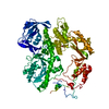
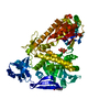
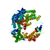
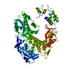
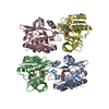
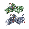
| Title | Crystal structure of the legionella pneumophila effector protein RavZ_20-502 | ||||||
 Components Components | Legionella pneumophila effector protein RavZ | ||||||
 Keywords Keywords | HYDROLASE / Hydrolase / Autophagy / Legionella pneumophila effector protein / ATG8 deconjugating enzyme | ||||||
| Function / homology |  Function and homology information Function and homology informationhost intracellular membrane-bounded organelle / protein delipidation / symbiont-mediated suppression of host autophagy / phosphatidylinositol-3-phosphate binding / cysteine-type peptidase activity / host cell cytoplasmic vesicle membrane / Hydrolases; Acting on peptide bonds (peptidases); Cysteine endopeptidases / proteolysis / extracellular region Similarity search - Function | ||||||
| Biological species |  Legionella pneumophila subsp. pneumophila ATCC 33215 (bacteria) Legionella pneumophila subsp. pneumophila ATCC 33215 (bacteria) | ||||||
| Method |  X-RAY DIFFRACTION / SYNCHROTRON / MOLECULAR REPLACEMENT / Resolution: 2.8 Å X-RAY DIFFRACTION / SYNCHROTRON / MOLECULAR REPLACEMENT / Resolution: 2.8 Å | ||||||
 Authors Authors | Pantoom, S. / Vetter, I.R. / Wu, Y.W. | ||||||
 Citation Citation | Journal: Elife / Year: 2017 Title: Elucidation of the anti-autophagy mechanism of the Legionella effector RavZ using semisynthetic LC3 proteins. Authors: Yang, A. / Pantoom, S. / Wu, Y.W. | ||||||
| History |
|
- Structure visualization
| Structure viewer | Molecule: MolmilJmol/JSmol |
|---|
- Downloads & links
-Download
| PDBx/mmCIF format | 5ms7.cif.gz | 168.5 KB | Display | PDBx/mmCIF format |
|---|---|---|---|---|
| PDB format | pdb5ms7.ent.gz | 133.2 KB | Display | PDB format |
| PDBx/mmJSON format | 5ms7.json.gz | Tree view | PDBx/mmJSON format | |
| Others |  Other downloads Other downloads |
-Validation report
| Arichive directory | https://data.pdbj.org/pub/pdb/validation_reports/ms/5ms7ftp://data.pdbj.org/pub/pdb/validation_reports/ms/5ms7 | HTTPS FTP |
|---|
-Related structure data
| Related structure data |  5ms2C  5ms5C  5ms6C 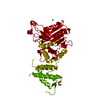 5ms8C 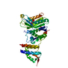 5cqcS S: Starting model for refinement C: citing same article ( |
|---|---|
| Similar structure data |

-Links
 PDBj
PDBj- Assembly
| Deposited unit | 
| ||||||||
|---|---|---|---|---|---|---|---|---|---|
| 1 | 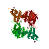
| ||||||||
| Unit cell |
| ||||||||
| Components on special symmetry positions |
|
-Components
| #1: Protein | Mass: 54285.918 Da / Num. of mol.: 1 Source method: isolated from a genetically manipulated source Source: (gene. exp.) Legionella pneumophila subsp. pneumophila ATCC 33215 (bacteria)Gene: lpg1683 / Plasmid: pOPIN / Production host: | ||||
|---|---|---|---|---|---|
| #2: Chemical |   Mass: 137.327 Da / Num. of mol.: 2 / Source method: obtained synthetically / Formula: Ba Mass: 137.327 Da / Num. of mol.: 2 / Source method: obtained synthetically / Formula: Ba#3: Chemical |   Mass: 92.094 Da / Num. of mol.: 3 / Source method: obtained synthetically / Formula: C3H8O3 Mass: 92.094 Da / Num. of mol.: 3 / Source method: obtained synthetically / Formula: C3H8O3#4: Water | ChemComp-HOH / |  Mass: 18.015 Da / Num. of mol.: 14 / Source method: isolated from a natural source / Formula: H2O Mass: 18.015 Da / Num. of mol.: 14 / Source method: isolated from a natural source / Formula: H2O |
-Experimental details
-Experiment
| Experiment | Method: X-RAY DIFFRACTION / Number of used crystals: 1 |
|---|
- Sample preparation
| Crystal | Density Matthews: 4.17 Å3/Da / Density % sol: 70.49 % |
|---|---|
| Crystal grow | Temperature: 277.15 K / Method: vapor diffusion, hanging drop / pH: 5.6 / Details: 16% PEG3350, 0.2 M BaCl2 and 0.1 M MES |
-Data collection
| Diffraction | Mean temperature: 100 K |
|---|---|
| Diffraction source | Source: SYNCHROTRON / Site: SLS  / Beamline: X10SA / Wavelength: 0.97862 Å / Beamline: X10SA / Wavelength: 0.97862 Å |
| Detector | Type: PSI PILATUS 6M / Detector: PIXEL / Date: Feb 26, 2016 |
| Radiation | Monochromator: SI (111) / Protocol: SINGLE WAVELENGTH / Monochromatic (M) / Laue (L): M / Scattering type: x-ray |
| Radiation wavelength | Wavelength: 0.97862 Å / Relative weight: 1 |
| Reflection | Resolution: 2.8→47.15 Å / Num. obs: 22921 / % possible obs: 100 % / Redundancy: 26.7 % / Biso Wilson estimate: 86.3 Å2 / CC1/2: 0.999 / Rmerge(I) obs: 0.1938 / Net I/σ(I): 15.14 |
| Reflection shell | Resolution: 2.8→2.9 Å / Redundancy: 26.3 % / Rmerge(I) obs: 1.951 / Mean I/σ(I) obs: 1.94 / CC1/2: 0.165 / % possible all: 100 |
- Processing
| Software |
| ||||||||||||||||||||||||||||||||||||||||||||||||||||||||||||||||||||||||||||||||||||||||||||||||||||
|---|---|---|---|---|---|---|---|---|---|---|---|---|---|---|---|---|---|---|---|---|---|---|---|---|---|---|---|---|---|---|---|---|---|---|---|---|---|---|---|---|---|---|---|---|---|---|---|---|---|---|---|---|---|---|---|---|---|---|---|---|---|---|---|---|---|---|---|---|---|---|---|---|---|---|---|---|---|---|---|---|---|---|---|---|---|---|---|---|---|---|---|---|---|---|---|---|---|---|---|---|---|
| Refinement | Method to determine structure: MOLECULAR REPLACEMENT Starting model: 5CQC Resolution: 2.8→47.145 Å / SU ML: 0.48 / Cross valid method: NONE / σ(F): 1.37 / Phase error: 30.74
| ||||||||||||||||||||||||||||||||||||||||||||||||||||||||||||||||||||||||||||||||||||||||||||||||||||
| Solvent computation | Shrinkage radii: 0.9 Å / VDW probe radii: 1.11 Å | ||||||||||||||||||||||||||||||||||||||||||||||||||||||||||||||||||||||||||||||||||||||||||||||||||||
| Refinement step | Cycle: LAST / Resolution: 2.8→47.145 Å
| ||||||||||||||||||||||||||||||||||||||||||||||||||||||||||||||||||||||||||||||||||||||||||||||||||||
| Refine LS restraints |
| ||||||||||||||||||||||||||||||||||||||||||||||||||||||||||||||||||||||||||||||||||||||||||||||||||||
| LS refinement shell |
| ||||||||||||||||||||||||||||||||||||||||||||||||||||||||||||||||||||||||||||||||||||||||||||||||||||
| Refinement TLS params. | Method: refined / Refine-ID: X-RAY DIFFRACTION
| ||||||||||||||||||||||||||||||||||||||||||||||||||||||||||||||||||||||||||||||||||||||||||||||||||||
| Refinement TLS group |
|
