 Movie
Movie Controller


+ Open data

- Basic information
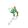
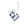
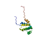
| Entry | Database: PDB / ID: 5ms5 | ||||||
|---|---|---|---|---|---|---|---|
| Title | Low-salt structure of RavZ LIR2-fused human LC3B | ||||||
 Components Components | RavZ,Microtubule-associated proteins 1A/1B light chain 3B | ||||||
 Keywords Keywords | PROTEIN BINDING / Autophagy / Host-pathogen interaction / Protein binding /RavZ / LIR / LC3 | ||||||
| Function / homology |  Function and homology information Function and homology informationhost intracellular membrane-bounded organelle / protein delipidation / SARS-CoV-2 modulates autophagy / ceramide binding / symbiont-mediated suppression of host autophagy / phosphatidylinositol-3-phosphate binding / phosphatidylethanolamine binding / Translation of Replicase and Assembly of the Replication Transcription Complex / TBC/RABGAPs / cellular response to nitrogen starvation ...host intracellular membrane-bounded organelle / protein delipidation / SARS-CoV-2 modulates autophagy / ceramide binding / symbiont-mediated suppression of host autophagy / phosphatidylinositol-3-phosphate binding / phosphatidylethanolamine binding / Translation of Replicase and Assembly of the Replication Transcription Complex / TBC/RABGAPs / cellular response to nitrogen starvation / Receptor Mediated Mitophagy / Macroautophagy / organelle membrane / autophagosome membrane / autophagosome maturation / axoneme / autophagosome assembly / mitophagy / cysteine-type peptidase activity / endomembrane system / autophagosome / Pexophagy / cellular response to starvation / PINK1-PRKN Mediated Mitophagy / macroautophagy / host cell cytoplasmic vesicle membrane / autophagy / mitochondrial membrane / KEAP1-NFE2L2 pathway / cytoplasmic vesicle / Translation of Replicase and Assembly of the Replication Transcription Complex / microtubule binding / microtubule / Hydrolases; Acting on peptide bonds (peptidases); Cysteine endopeptidases / ubiquitin protein ligase binding / mitochondrion / proteolysis / extracellular region / cytosol Similarity search - Function | ||||||
| Biological species |   Legionella pneumophila (bacteria) Legionella pneumophila (bacteria) Homo sapiens (human) Homo sapiens (human) | ||||||
| Method |  X-RAY DIFFRACTION / SYNCHROTRON / MOLECULAR REPLACEMENT / Resolution: 1.53 Å X-RAY DIFFRACTION / SYNCHROTRON / MOLECULAR REPLACEMENT / Resolution: 1.53 Å | ||||||
 Authors Authors | Pantoom, S. / Vetter, I.R. / Wu, Y.W. | ||||||
 Citation Citation | Journal: Elife / Year: 2017 Title: Elucidation of the anti-autophagy mechanism of the Legionella effector RavZ using semisynthetic LC3 proteins. Authors: Yang, A. / Pantoom, S. / Wu, Y.W. | ||||||
| History |
|
- Structure visualization
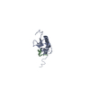
| Structure viewer | Molecule: MolmilJmol/JSmol |
|---|
- Downloads & links
-Download
| PDBx/mmCIF format | 5ms5.cif.gz | 123.3 KB | Display | PDBx/mmCIF format |
|---|---|---|---|---|
| PDB format | pdb5ms5.ent.gz | 97 KB | Display | PDB format |
| PDBx/mmJSON format | 5ms5.json.gz | Tree view | PDBx/mmJSON format | |
| Others |  Other downloads Other downloads |
-Validation report
| Arichive directory | https://data.pdbj.org/pub/pdb/validation_reports/ms/5ms5ftp://data.pdbj.org/pub/pdb/validation_reports/ms/5ms5 | HTTPS FTP |
|---|
-Related structure data
| Related structure data |  5ms2C  5ms6C  5ms7C 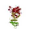 5ms8C  2z0eS S: Starting model for refinement C: citing same article ( |
|---|---|
| Similar structure data |


-Links
 PDBj
PDBj


- Assembly
| Deposited unit | 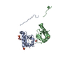
| ||||||||
|---|---|---|---|---|---|---|---|---|---|
| 1 | 
| ||||||||
| 2 | 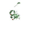
| ||||||||
| Unit cell |
| ||||||||
| Components on special symmetry positions |
|
-Components
| #1: Protein | Mass: 15968.087 Da / Num. of mol.: 2 Source method: isolated from a genetically manipulated source Details: The fusion protein of RavZ LIR (RESIDUES 25-36) and Microtubule-associated proteins 1A/1B light chain 3B (RESIDUES 2-119) Source: (gene. exp.) Legionella pneumophila (bacteria), (gene. exp.) Homo sapiens (human)Gene: lpg1683, MAP1LC3B, MAP1ALC3 / Plasmid: pMAL / Production host: #2: Chemical | ChemComp-SO4 /   Mass: 96.063 Da / Num. of mol.: 4 / Source method: obtained synthetically / Formula: SO4 Mass: 96.063 Da / Num. of mol.: 4 / Source method: obtained synthetically / Formula: SO4#3: Chemical | ChemComp-GOL / |   Mass: 92.094 Da / Num. of mol.: 1 / Source method: obtained synthetically / Formula: C3H8O3 Mass: 92.094 Da / Num. of mol.: 1 / Source method: obtained synthetically / Formula: C3H8O3#4: Water | ChemComp-HOH / |  Mass: 18.015 Da / Num. of mol.: 198 / Source method: isolated from a natural source / Formula: H2O Mass: 18.015 Da / Num. of mol.: 198 / Source method: isolated from a natural source / Formula: H2O |
|---|
-Experimental details
-Experiment
| Experiment | Method: X-RAY DIFFRACTION / Number of used crystals: 1 |
|---|
- Sample preparation
| Crystal | Density Matthews: 2.22 Å3/Da / Density % sol: 44.69 % |
|---|---|
| Crystal grow | Temperature: 293.15 K / Method: vapor diffusion, hanging drop Details: 0.1 M citric acid anhydrous and 1.6 M ammonium sulfate PH range: 3.6 |
-Data collection
| Diffraction | Mean temperature: 100 K |
|---|---|
| Diffraction source | Source: SYNCHROTRON / Site: SLS  / Beamline: X10SA / Wavelength: 0.99992 Å / Beamline: X10SA / Wavelength: 0.99992 Å |
| Detector | Type: DECTRIS PILATUS 6M / Detector: PIXEL / Date: Jun 16, 2016 |
| Radiation | Monochromator: SI (111) / Protocol: SINGLE WAVELENGTH / Monochromatic (M) / Laue (L): M / Scattering type: x-ray |
| Radiation wavelength | Wavelength: 0.99992 Å / Relative weight: 1 |
| Reflection | Resolution: 1.53→49.222 Å / Num. obs: 44250 / % possible obs: 100 % / Redundancy: 25.4 % / Biso Wilson estimate: 19.73 Å2 / CC1/2: 0.999 / Rmerge(I) obs: 0.1769 / Net I/σ(I): 14.69 |
| Reflection shell | Resolution: 1.53→1.585 Å / Redundancy: 24.6 % / Rmerge(I) obs: 2.205 / Mean I/σ(I) obs: 1.42 / CC1/2: 0.165 / % possible all: 100 |
- Processing
| Software |
| |||||||||||||||||||||||||||||||||||||||||||||||||||||||||||||||||||||||||||||||||||||||||||||||||||||||||||||||||||||||||||||||||||||||||||||||||||||||||||||||||||||||||||||||
|---|---|---|---|---|---|---|---|---|---|---|---|---|---|---|---|---|---|---|---|---|---|---|---|---|---|---|---|---|---|---|---|---|---|---|---|---|---|---|---|---|---|---|---|---|---|---|---|---|---|---|---|---|---|---|---|---|---|---|---|---|---|---|---|---|---|---|---|---|---|---|---|---|---|---|---|---|---|---|---|---|---|---|---|---|---|---|---|---|---|---|---|---|---|---|---|---|---|---|---|---|---|---|---|---|---|---|---|---|---|---|---|---|---|---|---|---|---|---|---|---|---|---|---|---|---|---|---|---|---|---|---|---|---|---|---|---|---|---|---|---|---|---|---|---|---|---|---|---|---|---|---|---|---|---|---|---|---|---|---|---|---|---|---|---|---|---|---|---|---|---|---|---|---|---|---|---|
| Refinement | Method to determine structure: MOLECULAR REPLACEMENT Starting model: 2Z0E Resolution: 1.53→49.222 Å / SU ML: 0.26 / Cross valid method: NONE / σ(F): 1.36 / Phase error: 28.35 / Stereochemistry target values: ML
| |||||||||||||||||||||||||||||||||||||||||||||||||||||||||||||||||||||||||||||||||||||||||||||||||||||||||||||||||||||||||||||||||||||||||||||||||||||||||||||||||||||||||||||||
| Solvent computation | Shrinkage radii: 0.9 Å / VDW probe radii: 1.11 Å / Solvent model: FLAT BULK SOLVENT MODEL | |||||||||||||||||||||||||||||||||||||||||||||||||||||||||||||||||||||||||||||||||||||||||||||||||||||||||||||||||||||||||||||||||||||||||||||||||||||||||||||||||||||||||||||||
| Refinement step | Cycle: LAST / Resolution: 1.53→49.222 Å
| |||||||||||||||||||||||||||||||||||||||||||||||||||||||||||||||||||||||||||||||||||||||||||||||||||||||||||||||||||||||||||||||||||||||||||||||||||||||||||||||||||||||||||||||
| Refine LS restraints |
| |||||||||||||||||||||||||||||||||||||||||||||||||||||||||||||||||||||||||||||||||||||||||||||||||||||||||||||||||||||||||||||||||||||||||||||||||||||||||||||||||||||||||||||||
| LS refinement shell |
| |||||||||||||||||||||||||||||||||||||||||||||||||||||||||||||||||||||||||||||||||||||||||||||||||||||||||||||||||||||||||||||||||||||||||||||||||||||||||||||||||||||||||||||||
| Refinement TLS params. | Method: refined / Refine-ID: X-RAY DIFFRACTION
| |||||||||||||||||||||||||||||||||||||||||||||||||||||||||||||||||||||||||||||||||||||||||||||||||||||||||||||||||||||||||||||||||||||||||||||||||||||||||||||||||||||||||||||||
| Refinement TLS group |
|
