 Movie
Movie Controller

[English] 日本語
 Yorodumi
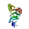
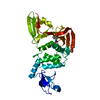
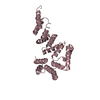
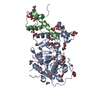
- PDB-5cqc: Crystal structure of the legionella pneumophila effector protein RavZ -
+ Open data

- Basic information
| Entry | Database: PDB / ID: 5cqc | ||||||
|---|---|---|---|---|---|---|---|
| Title | Crystal structure of the legionella pneumophila effector protein RavZ | ||||||
 Components Components | putative RavZ protein | ||||||
 Keywords Keywords | HYDROLASE / Ulp-family cysteine protease / autophagy inhibitor / PI3P binding domain / legionella pneumophila effector protein | ||||||
| Function / homology |  Function and homology information Function and homology informationhost intracellular membrane-bounded organelle / protein delipidation / symbiont-mediated suppression of host autophagy / phosphatidylinositol-3-phosphate binding / cysteine-type peptidase activity / host cell cytoplasmic vesicle membrane / Hydrolases; Acting on peptide bonds (peptidases); Cysteine endopeptidases / proteolysis / extracellular region Similarity search - Function | ||||||
| Biological species |   Legionella pneumophila (bacteria) Legionella pneumophila (bacteria) | ||||||
| Method |  X-RAY DIFFRACTION / SYNCHROTRON / MOLECULAR REPLACEMENT / Resolution: 2.985 Å X-RAY DIFFRACTION / SYNCHROTRON / MOLECULAR REPLACEMENT / Resolution: 2.985 Å | ||||||
 Authors Authors | Horenkamp, F.A. / Reinisch, K.M. | ||||||
 Citation Citation | Journal: Dev.Cell / Year: 2015 Title: The Legionella Anti-autophagy Effector RavZ Targets the Autophagosome via PI3P- and Curvature-Sensing Motifs. Authors: Horenkamp, F.A. / Kauffman, K.J. / Kohler, L.J. / Sherwood, R.K. / Krueger, K.P. / Shteyn, V. / Roy, C.R. / Melia, T.J. / Reinisch, K.M. | ||||||
| History |
|
- Structure visualization
| Structure viewer | Molecule: MolmilJmol/JSmol |
|---|
- Downloads & links
-Download
| PDBx/mmCIF format | 5cqc.cif.gz | 151.9 KB | Display | PDBx/mmCIF format |
|---|---|---|---|---|
| PDB format | pdb5cqc.ent.gz | 119 KB | Display | PDB format |
| PDBx/mmJSON format | 5cqc.json.gz | Tree view | PDBx/mmJSON format | |
| Others |  Other downloads Other downloads |
-Validation report
| Arichive directory | https://data.pdbj.org/pub/pdb/validation_reports/cq/5cqcftp://data.pdbj.org/pub/pdb/validation_reports/cq/5cqc | HTTPS FTP |
|---|
-Related structure data
| Similar structure data |
|---|


-Links
 PDBj
PDBj- Assembly
| Deposited unit | 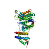
| ||||||||
|---|---|---|---|---|---|---|---|---|---|
| 1 |
| ||||||||
| Unit cell |
|
-Components
| #1: Protein | Mass: 47118.910 Da / Num. of mol.: 1 Source method: isolated from a genetically manipulated source Source: (gene. exp.) Legionella pneumophila (bacteria) / Gene: lpg1683 / Plasmid: pGex6P1 / Production host: |
|---|---|
| #2: Chemical | ChemComp-BA /   Mass: 137.327 Da / Num. of mol.: 1 / Source method: obtained synthetically / Formula: Ba Mass: 137.327 Da / Num. of mol.: 1 / Source method: obtained synthetically / Formula: Ba |
| #3: Water | ChemComp-HOH /  Mass: 18.015 Da / Num. of mol.: 1 / Source method: isolated from a natural source / Formula: H2O Mass: 18.015 Da / Num. of mol.: 1 / Source method: isolated from a natural source / Formula: H2O |
-Experimental details
-Experiment
| Experiment | Method: X-RAY DIFFRACTION |
|---|
- Sample preparation
| Crystal | Density Matthews: 4.73 Å3/Da / Density % sol: 74.01 % |
|---|---|
| Crystal grow | Temperature: 277.15 K / Method: vapor diffusion, hanging drop / pH: 5.1 / Details: 12% PEG 3350, 0.2 M BaCl2, 0.1 M MES |
-Data collection
| Diffraction | Mean temperature: 100 K |
|---|---|
| Diffraction source | Source: SYNCHROTRON / Site: APS  / Beamline: 24-ID-C / Wavelength: 0.9792 Å / Beamline: 24-ID-C / Wavelength: 0.9792 Å |
| Detector | Type: ADSC QUANTUM 315 / Detector: CCD / Date: Jul 9, 2014 |
| Radiation | Monochromator: Si(220) / Protocol: SINGLE WAVELENGTH / Monochromatic (M) / Laue (L): M / Scattering type: x-ray |
| Radiation wavelength | Wavelength: 0.9792 Å / Relative weight: 1 |
| Reflection | Resolution: 2.98→110 Å / Num. all: 18551 / Num. obs: 18194 / % possible obs: 99.2 % / Redundancy: 5.7 % / Rmerge(I) obs: 0.066 / Net I/σ(I): 17.3 |
| Reflection shell | Resolution: 2.98→3.14 Å / Redundancy: 5.9 % / Mean I/σ(I) obs: 0.9 / % possible all: 98.2 |
- Processing
| Software |
| ||||||||||||||||||||||||||||||||||||||||||||||||||||||||||||||||||||||||||||||||||||||||||||||||||||
|---|---|---|---|---|---|---|---|---|---|---|---|---|---|---|---|---|---|---|---|---|---|---|---|---|---|---|---|---|---|---|---|---|---|---|---|---|---|---|---|---|---|---|---|---|---|---|---|---|---|---|---|---|---|---|---|---|---|---|---|---|---|---|---|---|---|---|---|---|---|---|---|---|---|---|---|---|---|---|---|---|---|---|---|---|---|---|---|---|---|---|---|---|---|---|---|---|---|---|---|---|---|
| Refinement | Method to determine structure: MOLECULAR REPLACEMENT / Resolution: 2.985→68.942 Å / SU ML: 0.44 / Cross valid method: FREE R-VALUE / σ(F): 1.34 / Phase error: 29.61 / Stereochemistry target values: ML
| ||||||||||||||||||||||||||||||||||||||||||||||||||||||||||||||||||||||||||||||||||||||||||||||||||||
| Solvent computation | Shrinkage radii: 0.9 Å / VDW probe radii: 1.11 Å / Solvent model: FLAT BULK SOLVENT MODEL | ||||||||||||||||||||||||||||||||||||||||||||||||||||||||||||||||||||||||||||||||||||||||||||||||||||
| Refinement step | Cycle: LAST / Resolution: 2.985→68.942 Å
| ||||||||||||||||||||||||||||||||||||||||||||||||||||||||||||||||||||||||||||||||||||||||||||||||||||
| Refine LS restraints |
| ||||||||||||||||||||||||||||||||||||||||||||||||||||||||||||||||||||||||||||||||||||||||||||||||||||
| LS refinement shell |
| ||||||||||||||||||||||||||||||||||||||||||||||||||||||||||||||||||||||||||||||||||||||||||||||||||||
| Refinement TLS params. | Method: refined / Refine-ID: X-RAY DIFFRACTION
| ||||||||||||||||||||||||||||||||||||||||||||||||||||||||||||||||||||||||||||||||||||||||||||||||||||
| Refinement TLS group |
|
