 Movie
Movie Controller

[English] 日本語
 Yorodumi
- PDB-5xw7: Crystal structure of the flexible tandem repeat domain of bacteri... -
+ Open data

- Basic information
| Entry | Database: PDB / ID: 5xw7 | ||||||
|---|---|---|---|---|---|---|---|
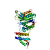
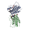
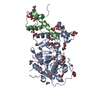
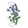
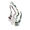
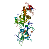
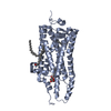
| Title | Crystal structure of the flexible tandem repeat domain of bacterial cellulose synthase subunit C | ||||||
 Components Components | Cellulose synthase subunit C | ||||||
 Keywords Keywords | BIOSYNTHETIC PROTEIN / cellulose synthase / tetratrico peptide repeat | ||||||
| Function / homology |  Function and homology information Function and homology information | ||||||
| Biological species |  Enterobacter sp. CJF-002 (bacteria) Enterobacter sp. CJF-002 (bacteria) | ||||||
| Method |  X-RAY DIFFRACTION / SYNCHROTRON / SAD / Resolution: 3.272 Å X-RAY DIFFRACTION / SYNCHROTRON / SAD / Resolution: 3.272 Å | ||||||
 Authors Authors | Nojima, S. / Kato, K. / Yao, M. | ||||||
 Citation Citation | Journal: Sci Rep / Year: 2017 Title: Crystal structure of the flexible tandem repeat domain of bacterial cellulose synthesis subunit C. Authors: Shingo Nojima / Ayumi Fujishima / Koji Kato / Kayoko Ohuchi / Nobutaka Shimizu / Kento Yonezawa / Kenji Tajima / Min Yao /  Abstract: Bacterial cellulose (BC) is synthesized and exported through the cell membrane via a large protein complex (terminal complex) that consists of three or four subunits. BcsC is a little-studied subunit ...Bacterial cellulose (BC) is synthesized and exported through the cell membrane via a large protein complex (terminal complex) that consists of three or four subunits. BcsC is a little-studied subunit considered to export BC to the extracellular matrix. It is predicted to have two domains: a tetratrico peptide repeat (TPR) domain and a β-barrelled outer membrane domain. Here we report the crystal structure of the N-terminal part of BcsC-TPR domain (Asp24-Arg272) derived from Enterobacter CJF-002. Unlike most TPR-containing proteins which have continuous TPR motifs, this structure has an extra α-helix between two clusters of TPR motifs. Five independent molecules in the crystal had three different conformations that varied at the hinge of the inserted α-helix. Such structural feature indicates that the inserted α-helix confers flexibility to the chain and changes the direction of the TPR super-helix, which was also suggested by structural analysis of BcsC-TPR (Asp24-Leu664) in solution by size exclusion chromatography-small-angle X-ray scattering. The flexibility at the α-helical hinge may play important role for exporting glucan chains. | ||||||
| History |
|
- Structure visualization
| Structure viewer | Molecule: MolmilJmol/JSmol |
|---|
- Downloads & links
-Download
| PDBx/mmCIF format | 5xw7.cif.gz | 219.6 KB | Display | PDBx/mmCIF format |
|---|---|---|---|---|
| PDB format | pdb5xw7.ent.gz | 179 KB | Display | PDB format |
| PDBx/mmJSON format | 5xw7.json.gz | Tree view | PDBx/mmJSON format | |
| Others |  Other downloads Other downloads |
-Validation report
| Arichive directory | https://data.pdbj.org/pub/pdb/validation_reports/xw/5xw7ftp://data.pdbj.org/pub/pdb/validation_reports/xw/5xw7 | HTTPS FTP |
|---|
-Related structure data
| Related structure data | C: citing same article ( |
|---|---|
| Similar structure data |


-Links
 PDBj
PDBj


- Assembly
| Deposited unit | 
| ||||||||
|---|---|---|---|---|---|---|---|---|---|
| 1 | 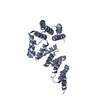
| ||||||||
| 2 | 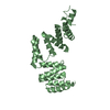
| ||||||||
| 3 | 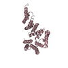
| ||||||||
| 4 | 
| ||||||||
| 5 | 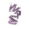
| ||||||||
| Unit cell |
|
-Components
| #1: Protein | Mass: 28554.031 Da / Num. of mol.: 5 / Fragment: UNP residues 24-272 Source method: isolated from a genetically manipulated source Details: chainB: S230A, G231A, D232A, T233A, V234A, D235A, S236A, R238A, T239A, Q240A Source: (gene. exp.) Enterobacter sp. CJF-002 (bacteria) / Gene: bcsC / Production host: |
|---|
-Experimental details
-Experiment
| Experiment | Method: X-RAY DIFFRACTION / Number of used crystals: 1 |
|---|
- Sample preparation
| Crystal | Density Matthews: 3.71 Å3/Da / Density % sol: 66.8 % |
|---|---|
| Crystal grow | Temperature: 293 K / Method: vapor diffusion, sitting drop / Details: MES, sodium chloride |
-Data collection
| Diffraction |
| |||||||||||||||
|---|---|---|---|---|---|---|---|---|---|---|---|---|---|---|---|---|
| Diffraction source |
| |||||||||||||||
| Detector |
| |||||||||||||||
| Radiation |
| |||||||||||||||
| Radiation wavelength |
| |||||||||||||||
| Reflection | Resolution: 3.28→50 Å / Num. obs: 32542 / % possible obs: 98.6 % / Redundancy: 3.7 % / Net I/σ(I): 10.7 | |||||||||||||||
| Reflection shell | Resolution: 3.27→3.47 Å / Redundancy: 3.8 % / Rmerge(I) obs: 0.69 / Rsym value: 0.803 / % possible all: 98.2 |
- Processing
| Software |
| |||||||||||||||||||||||||||||||||||||||||||||||||||||||||||||||||||||||||||||||||||||||||||||||||||||||||||||||||||||||
|---|---|---|---|---|---|---|---|---|---|---|---|---|---|---|---|---|---|---|---|---|---|---|---|---|---|---|---|---|---|---|---|---|---|---|---|---|---|---|---|---|---|---|---|---|---|---|---|---|---|---|---|---|---|---|---|---|---|---|---|---|---|---|---|---|---|---|---|---|---|---|---|---|---|---|---|---|---|---|---|---|---|---|---|---|---|---|---|---|---|---|---|---|---|---|---|---|---|---|---|---|---|---|---|---|---|---|---|---|---|---|---|---|---|---|---|---|---|---|---|---|
| Refinement | Method to determine structure: SAD / Resolution: 3.272→47.695 Å / SU ML: 0.44 / Cross valid method: FREE R-VALUE / σ(F): 1.36 / Phase error: 26.88
| |||||||||||||||||||||||||||||||||||||||||||||||||||||||||||||||||||||||||||||||||||||||||||||||||||||||||||||||||||||||
| Solvent computation | Shrinkage radii: 0.9 Å / VDW probe radii: 1.11 Å | |||||||||||||||||||||||||||||||||||||||||||||||||||||||||||||||||||||||||||||||||||||||||||||||||||||||||||||||||||||||
| Refinement step | Cycle: LAST / Resolution: 3.272→47.695 Å
| |||||||||||||||||||||||||||||||||||||||||||||||||||||||||||||||||||||||||||||||||||||||||||||||||||||||||||||||||||||||
| Refine LS restraints |
| |||||||||||||||||||||||||||||||||||||||||||||||||||||||||||||||||||||||||||||||||||||||||||||||||||||||||||||||||||||||
| LS refinement shell |
|
