 ムービー
ムービー コントローラー


+ データを開く

- 基本情報
基本情報
| 登録情報 | データベース: PDB / ID: 5hzv | ||||||||||||||||||||||||
|---|---|---|---|---|---|---|---|---|---|---|---|---|---|---|---|---|---|---|---|---|---|---|---|---|---|
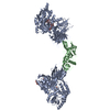
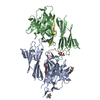
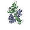
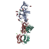
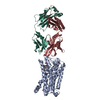
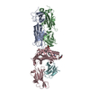
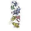
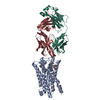
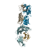
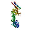
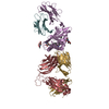
| タイトル | Crystal structure of the zona pellucida module of human endoglin/CD105 | ||||||||||||||||||||||||
 要素 要素 | Maltose-binding periplasmic protein,Endoglin | ||||||||||||||||||||||||
 キーワード キーワード | SIGNALING PROTEIN / ZONA PELLUCIDA DOMAIN / ANGIOGENESIS / GLYCOPROTEIN / RECEPTOR | ||||||||||||||||||||||||
| 機能・相同性 |  機能・相同性情報 機能・相同性情報atrioventricular canal morphogenesis / detection of hypoxia / endothelial microparticle / venous blood vessel morphogenesis / dorsal aorta morphogenesis / positive regulation of vascular associated smooth muscle cell differentiation / cell migration involved in endocardial cushion formation / vascular associated smooth muscle cell development / atrial cardiac muscle tissue morphogenesis / central nervous system vasculogenesis ...atrioventricular canal morphogenesis / detection of hypoxia / endothelial microparticle / venous blood vessel morphogenesis / dorsal aorta morphogenesis / positive regulation of vascular associated smooth muscle cell differentiation / cell migration involved in endocardial cushion formation / vascular associated smooth muscle cell development / atrial cardiac muscle tissue morphogenesis / central nervous system vasculogenesis / epithelial to mesenchymal transition involved in endocardial cushion formation / positive regulation of epithelial to mesenchymal transition involved in endocardial cushion formation / cardiac ventricle morphogenesis / regulation of transforming growth factor beta receptor signaling pathway / galactose binding / cardiac atrium morphogenesis / smooth muscle tissue development / type II transforming growth factor beta receptor binding / activin binding / regulation of phosphorylation / type I transforming growth factor beta receptor binding / ventricular trabecula myocardium morphogenesis / glycosaminoglycan binding / positive regulation of BMP signaling pathway / outflow tract septum morphogenesis / transforming growth factor beta binding / artery morphogenesis / endocardial cushion morphogenesis / branching involved in blood vessel morphogenesis / negative regulation of SMAD protein signal transduction / detection of maltose stimulus / heart looping / maltose transport complex / negative regulation of endothelial cell proliferation / positive regulation of systemic arterial blood pressure / carbohydrate transport / signaling receptor activator activity / positive regulation of SMAD protein signal transduction / extracellular matrix disassembly / carbohydrate transmembrane transporter activity / maltose binding / maltose transport / maltodextrin transmembrane transport / BMP signaling pathway / vasculogenesis / regulation of cell adhesion / coreceptor activity / ATP-binding cassette (ABC) transporter complex, substrate-binding subunit-containing / ATP-binding cassette (ABC) transporter complex / transforming growth factor beta receptor signaling pathway / negative regulation of cell migration / cell chemotaxis / cell motility / negative regulation of transforming growth factor beta receptor signaling pathway / wound healing / positive regulation of angiogenesis / transmembrane signaling receptor activity / cell migration / regulation of cell population proliferation / outer membrane-bounded periplasmic space / periplasmic space / response to hypoxia / receptor complex / positive regulation of phosphatidylinositol 3-kinase/protein kinase B signal transduction / cell adhesion / nuclear body / negative regulation of gene expression / external side of plasma membrane / focal adhesion / DNA damage response / regulation of DNA-templated transcription / cell surface / negative regulation of transcription by RNA polymerase II / protein homodimerization activity / positive regulation of transcription by RNA polymerase II / extracellular space / identical protein binding / membrane / plasma membrane 類似検索 - 分子機能 | ||||||||||||||||||||||||
| 生物種 |   Homo sapiens (ヒト) Homo sapiens (ヒト) | ||||||||||||||||||||||||
| 手法 |  X線回折 / シンクロトロン / 分子置換 / 解像度: 2.7 Å X線回折 / シンクロトロン / 分子置換 / 解像度: 2.7 Å | ||||||||||||||||||||||||
 データ登録者 データ登録者 | Bokhove, M. / Saito, T. / Jovine, L. | ||||||||||||||||||||||||
| 資金援助 |  スウェーデン, 7件 スウェーデン, 7件
| ||||||||||||||||||||||||
 引用 引用 | ジャーナル: Cell Rep / 年: 2017 タイトル: Structural Basis of the Human Endoglin-BMP9 Interaction: Insights into BMP Signaling and HHT1. 著者: Saito, T. / Bokhove, M. / Croci, R. / Zamora-Caballero, S. / Han, L. / Letarte, M. / de Sanctis, D. / Jovine, L. #1: ジャーナル: J. Biol. Chem. / 年: 1990 タイトル: Primary structure of endoglin, an RGD-containing glycoprotein of human endothelial cells. 著者: Gougos, A. / Letarte, M. #2: ジャーナル: FEBS Lett. / 年: 1992 タイトル: A large domain common to sperm receptors (Zp2 and Zp3) and TGF-beta type III receptor. 著者: Bork, P. / Sander, C. #3: ジャーナル: Annu Rev Biochem / 年: 2005 タイトル: Zona pellucida domain proteins. 著者: Luca Jovine / Costel C Darie / Eveline S Litscher / Paul M Wassarman /  要旨: Many eukaryotic proteins share a sequence designated as the zona pellucida (ZP) domain. This structural element, present in extracellular proteins from a wide variety of organisms, from nematodes to ...Many eukaryotic proteins share a sequence designated as the zona pellucida (ZP) domain. This structural element, present in extracellular proteins from a wide variety of organisms, from nematodes to mammals, consists of approximately 260 amino acids with eight conserved cysteine (Cys) residues and is located close to the C terminus of the polypeptide. ZP domain proteins are often glycosylated, modular structures consisting of multiple types of domains. Predictions can be made about some of the structural features of the ZP domain and ZP domain proteins. The functions of ZP domain proteins vary tremendously, from serving as structural components of egg coats, appendicularian mucous houses, and nematode dauer larvae, to serving as mechanotransducers in flies and receptors in mammals and nonmammals. Generally, ZP domain proteins are present in filaments and/or matrices, which is consistent with the role of the domain in protein polymerization. A general mechanism for assembly of ZP domain proteins has been presented. It is likely that the ZP domain plays a common role despite its presence in proteins of widely diverse functions. #4: ジャーナル: Cell / 年: 2010タイトル: Insights into egg coat assembly and egg-sperm interaction from the X-ray structure of full-length ZP3. 著者: Ling Han / Magnus Monné / Hiroki Okumura / Thomas Schwend / Amy L Cherry / David Flot / Tsukasa Matsuda / Luca Jovine / 要旨: ZP3, a major component of the zona pellucida (ZP) matrix coating mammalian eggs, is essential for fertilization by acting as sperm receptor. By retaining a propeptide that contains a polymerization- ...ZP3, a major component of the zona pellucida (ZP) matrix coating mammalian eggs, is essential for fertilization by acting as sperm receptor. By retaining a propeptide that contains a polymerization-blocking external hydrophobic patch (EHP), we determined the crystal structure of an avian homolog of ZP3 at 2.0 Å resolution. The structure unveils the fold of a complete ZP domain module in a homodimeric arrangement required for secretion and reveals how EHP prevents premature incorporation of ZP3 into the ZP. This suggests mechanisms underlying polymerization and how local structural differences, reflected by alternative disulfide patterns, control the specificity of ZP subunit interaction. Close relative positioning of a conserved O-glycan important for sperm binding and the hypervariable, positively selected C-terminal region of ZP3 suggests a concerted role in the regulation of species-restricted gamete recognition. Alternative conformations of the area around the O-glycan indicate how sperm binding could trigger downstream events via intramolecular signaling. #5: ジャーナル: Proc Natl Acad Sci U S A / 年: 2016タイトル: A structured interdomain linker directs self-polymerization of human uromodulin. 著者: Marcel Bokhove / Kaoru Nishimura / Martina Brunati / Ling Han / Daniele de Sanctis / Luca Rampoldi / Luca Jovine /   要旨: Uromodulin (UMOD)/Tamm-Horsfall protein, the most abundant human urinary protein, plays a key role in chronic kidney diseases and is a promising therapeutic target for hypertension. Via its bipartite ...Uromodulin (UMOD)/Tamm-Horsfall protein, the most abundant human urinary protein, plays a key role in chronic kidney diseases and is a promising therapeutic target for hypertension. Via its bipartite zona pellucida module (ZP-N/ZP-C), UMOD forms extracellular filaments that regulate kidney electrolyte balance and innate immunity, as well as protect against renal stones. Moreover, salt-dependent aggregation of UMOD filaments in the urine generates a soluble molecular net that captures uropathogenic bacteria and facilitates their clearance. Despite the functional importance of its homopolymers, no structural information is available on UMOD and how it self-assembles into filaments. Here, we report the crystal structures of polymerization regions of human UMOD and mouse ZP2, an essential sperm receptor protein that is structurally related to UMOD but forms heteropolymers. The structure of UMOD reveals that an extensive hydrophobic interface mediates ZP-N domain homodimerization. This arrangement is required for filament formation and is directed by an ordered ZP-N/ZP-C linker that is not observed in ZP2 but is conserved in the sequence of deafness/Crohn's disease-associated homopolymeric glycoproteins α-tectorin (TECTA) and glycoprotein 2 (GP2). Our data provide an example of how interdomain linker plasticity can modulate the function of structurally similar multidomain proteins. Moreover, the architecture of UMOD rationalizes numerous pathogenic mutations in both UMOD and TECTA genes. | ||||||||||||||||||||||||
| 履歴 |
|
- 構造の表示
構造の表示
| 構造ビューア | 分子: MolmilJmol/JSmol |
|---|
- ダウンロードとリンク
-ダウンロード
| PDBx/mmCIF形式 | 5hzv.cif.gz | 249 KB | 表示 | PDBx/mmCIF形式 |
|---|---|---|---|---|
| PDB形式 | pdb5hzv.ent.gz | 200.2 KB | 表示 | PDB形式 |
| PDBx/mmJSON形式 | 5hzv.json.gz | ツリー表示 | PDBx/mmJSON形式 | |
| その他 |  その他のダウンロード その他のダウンロード |
-検証レポート
| 文書・要旨 | 5hzv_validation.pdf.gz | 805.9 KB | 表示 | wwPDB検証レポート |
|---|---|---|---|---|
| 文書・詳細版 | 5hzv_full_validation.pdf.gz | 808.5 KB | 表示 | |
| XML形式データ | 5hzv_validation.xml.gz | 21.6 KB | 表示 | |
| CIF形式データ | 5hzv_validation.cif.gz | 29.2 KB | 表示 | |
| アーカイブディレクトリ | https://data.pdbj.org/pub/pdb/validation_reports/hz/5hzvftp://data.pdbj.org/pub/pdb/validation_reports/hz/5hzv | HTTPS FTP |
-関連構造データ


-リンク
 PDBj
PDBj


- 集合体
集合体
| 登録構造単位 | 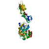
| ||||||||
|---|---|---|---|---|---|---|---|---|---|
| 1 |
| ||||||||
| 単位格子 |
|
-要素
| #1: タンパク質 | 分子量: 68228.398 Da / 分子数: 1 変異: D449A, K450A, E539A, N540A, A582H, K586H, K606A, A679V, I684V, E726A, E729A, D730A AND R734N,D449A, K450A, E539A, N540A, A582H, K586H, K606A, A679V, I684V, E726A, E729A, D730A AND ...変異: D449A, K450A, E539A, N540A, A582H, K586H, K606A, A679V, I684V, E726A, E729A, D730A AND R734N,D449A, K450A, E539A, N540A, A582H, K586H, K606A, A679V, I684V, E726A, E729A, D730A AND R734N,D449A, K450A, E539A, N540A, A582H, K586H, K606A, A679V, I684V, E726A, E729A, D730A AND R734N,D449A, K450A, E539A, N540A, A582H, K586H, K606A, A679V, I684V, E726A, E729A, D730A AND R734N,D449A, K450A, E539A, N540A, A582H, K586H, K606A, A679V, I684V, E726A, E729A, D730A AND R734N,D449A, K450A, E539A, N540A, A582H, K586H, K606A, A679V, I684V, E726A, E729A, D730A AND R734N,D449A, K450A, E539A, N540A, A582H, K586H, K606A, A679V, I684V, E726A, E729A, D730A AND R734N,D449A, K450A, E539A, N540A, A582H, K586H, K606A, A679V, I684V, E726A, E729A, D730A AND R734N,D449A, K450A, E539A, N540A, A582H, K586H, K606A, A679V, I684V, E726A, E729A, D730A AND R734N,D449A, K450A, E539A, N540A, A582H, K586H, K606A, A679V, I684V, E726A, E729A, D730A AND R734N,D449A, K450A, E539A, N540A, A582H, K586H, K606A, A679V, I684V, E726A, E729A, D730A AND R734N,D449A, K450A, E539A, N540A, A582H, K586H, K606A, A679V, I684V, E726A, E729A, D730A AND R734N,D449A, K450A, E539A, N540A, A582H, K586H, K606A, A679V, I684V, E726A, E729A, D730A AND R734N,D449A, K450A, E539A, N540A, A582H, K586H, K606A, A679V, I684V, E726A, E729A, D730A AND R734N,D449A, K450A, E539A, N540A, A582H, K586H, K606A, A679V, I684V, E726A, E729A, D730A AND R734N,D449A, K450A, E539A, N540A, A582H, K586H, K606A, A679V, I684V, E726A, E729A, D730A AND R734N 由来タイプ: 組換発現 詳細: THIS PROTEIN IS A CHIMERA. RESIDUES 368-734 ARE FROM E. COLI MALTOSE BINDING PROTEIN (MBP), CORRESPOND TO RESIDUES 27-393 OF SWISS-PROT DATABASE ENTRY P0AEX9 AND CONTAIN MUTATIONS D449A, ...詳細: THIS PROTEIN IS A CHIMERA. RESIDUES 368-734 ARE FROM E. COLI MALTOSE BINDING PROTEIN (MBP), CORRESPOND TO RESIDUES 27-393 OF SWISS-PROT DATABASE ENTRY P0AEX9 AND CONTAIN MUTATIONS D449A, K450A, E539A, N540A, A582H, K586H, K606A, A679V, I684V, E726A, E729A, D730A AND R734N (CORRESPONDING TO D108A, K109A, E198A, N199A, A241H, K245H, K265A, A338V, I343V, E385A, E388A, D389A AND R393N IN P0AEX9). RESIDUES 738-981 ARE FROM HUMAN ENDOGLIN PROTEIN AND CORRESPOND TO RESIDUES 338-581 OF SWISS-PROT DATABASE ENTRY P17813. SUBTRACTING 400 FROM THE PDB ENTRY RESIDUE NUMBERING RESULTS IN THE NUMBERING ACCORDING TO UNIPROT ENTRY P17813. 由来: (組換発現) Homo sapiens (ヒト)株: K12 / Cell: ENDOTHELIAL / 遺伝子: malE, b4034, JW3994, ENG, END / プラスミド: pHLsec / 細胞株 (発現宿主): HEK293T / 発現宿主: Homo sapiens (ヒト) / 参照: UniProt: P0AEX9, UniProt: P17813 |
|---|---|
| #2: 多糖 | alpha-D-glucopyranose-(1-4)-alpha-D-glucopyranose / alpha-maltose  |
| #3: 化合物 | ChemComp-GOL /   分子量: 92.094 Da / 分子数: 1 / 由来タイプ: 合成 / 式: C3H8O3 分子量: 92.094 Da / 分子数: 1 / 由来タイプ: 合成 / 式: C3H8O3 |
| #4: 水 | ChemComp-HOH /  分子量: 18.015 Da / 分子数: 15 / 由来タイプ: 天然 / 式: H2O 分子量: 18.015 Da / 分子数: 15 / 由来タイプ: 天然 / 式: H2O |
| Has protein modification | Y |
-実験情報
-実験
| 実験 | 手法: X線回折 / 使用した結晶の数: 1 |
|---|
- 試料調製
試料調製
| 結晶 | マシュー密度: 2.94 Å3/Da / 溶媒含有率: 58.2 % / 解説: Hexagonal rod |
|---|---|
| 結晶化 | 温度: 293 K / 手法: 蒸気拡散法, ハンギングドロップ法 / pH: 6.5 詳細: 11.5% equal mixture of MPD, PEG 1000 and PEG 3350 (1:1:1), MES/imidazole mix PH範囲: 5.5-7.0 |
-データ収集
| 回折 | 平均測定温度: 100 K |
|---|---|
| 放射光源 | 由来: シンクロトロン / サイト: ESRF / ビームライン: ID23-1 / 波長: 1 Å |
| 検出器 | タイプ: PSI PILATUS 6M / 検出器: PIXEL / 日付: 2015年9月25日 |
| 放射 | モノクロメーター: Si Single Crystal / プロトコル: SINGLE WAVELENGTH / 単色(M)・ラウエ(L): M / 散乱光タイプ: x-ray |
| 放射波長 | 波長: 1 Å / 相対比: 1 |
| 反射 | 解像度: 2.69→29.51 Å / Num. obs: 21583 / % possible obs: 97.9 % / Observed criterion σ(I): -3 / 冗長度: 3.3 % / Biso Wilson estimate: 72.174 Å2 / Rsym value: 0.062 / Net I/σ(I): 9.43 |
| 反射 シェル | 解像度: 2.69→2.76 Å / 冗長度: 2.8 % / Rmerge(I) obs: 0.675 / Mean I/σ(I) obs: 0.99 / % possible all: 85.4 |
- 解析
解析
| ソフトウェア |
| ||||||||||||||||||||||||||||||||||||||||||||||||||||||||||||||||||||||||||||||||||||||||||||||||||||||||
|---|---|---|---|---|---|---|---|---|---|---|---|---|---|---|---|---|---|---|---|---|---|---|---|---|---|---|---|---|---|---|---|---|---|---|---|---|---|---|---|---|---|---|---|---|---|---|---|---|---|---|---|---|---|---|---|---|---|---|---|---|---|---|---|---|---|---|---|---|---|---|---|---|---|---|---|---|---|---|---|---|---|---|---|---|---|---|---|---|---|---|---|---|---|---|---|---|---|---|---|---|---|---|---|---|---|
| 精密化 | 構造決定の手法: 分子置換 開始モデル: 3SEX 解像度: 2.7→29.5037 Å / SU ML: 0.51 / 交差検証法: FREE R-VALUE / σ(F): 1.33 / 位相誤差: 36.22 / 立体化学のターゲット値: ML
| ||||||||||||||||||||||||||||||||||||||||||||||||||||||||||||||||||||||||||||||||||||||||||||||||||||||||
| 溶媒の処理 | 減衰半径: 0.9 Å / VDWプローブ半径: 1.11 Å / 溶媒モデル: FLAT BULK SOLVENT MODEL | ||||||||||||||||||||||||||||||||||||||||||||||||||||||||||||||||||||||||||||||||||||||||||||||||||||||||
| 精密化ステップ | サイクル: LAST / 解像度: 2.7→29.5037 Å
| ||||||||||||||||||||||||||||||||||||||||||||||||||||||||||||||||||||||||||||||||||||||||||||||||||||||||
| 拘束条件 |
| ||||||||||||||||||||||||||||||||||||||||||||||||||||||||||||||||||||||||||||||||||||||||||||||||||||||||
| LS精密化 シェル | Refine-ID: X-RAY DIFFRACTION
| ||||||||||||||||||||||||||||||||||||||||||||||||||||||||||||||||||||||||||||||||||||||||||||||||||||||||
| 精密化 TLS | 手法: refined / Refine-ID: X-RAY DIFFRACTION
| ||||||||||||||||||||||||||||||||||||||||||||||||||||||||||||||||||||||||||||||||||||||||||||||||||||||||
| 精密化 TLSグループ |
|
