 Movie
Movie Controller

[English] 日本語
 Yorodumi
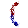
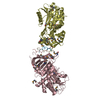
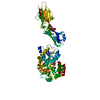
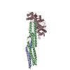
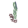
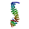
- PDB-2hft: THE CRYSTAL STRUCTURE OF THE EXTRACELLULAR DOMAIN OF HUMAN TISSUE... -
+ Open data

- Basic information
| Entry | Database: PDB / ID: 2hft | |||||||||
|---|---|---|---|---|---|---|---|---|---|---|
| Title | THE CRYSTAL STRUCTURE OF THE EXTRACELLULAR DOMAIN OF HUMAN TISSUE FACTOR AT 1.7 ANGSTROMS RESOLUTION | |||||||||
 Components Components | HUMAN TISSUE FACTOR | |||||||||
 Keywords Keywords | COAGULATION FACTOR | |||||||||
| Function / homology |  Function and homology information Function and homology informationactivation of plasma proteins involved in acute inflammatory response / activation of blood coagulation via clotting cascade / serine-type peptidase complex / positive regulation of platelet-derived growth factor receptor signaling pathway / NGF-stimulated transcription / cytokine receptor activity / positive regulation of positive chemotaxis / : / positive regulation of endothelial cell apoptotic process / positive regulation of TOR signaling ...activation of plasma proteins involved in acute inflammatory response / activation of blood coagulation via clotting cascade / serine-type peptidase complex / positive regulation of platelet-derived growth factor receptor signaling pathway / NGF-stimulated transcription / cytokine receptor activity / positive regulation of positive chemotaxis / : / positive regulation of endothelial cell apoptotic process / positive regulation of TOR signaling / positive regulation of endothelial cell proliferation / positive regulation of interleukin-8 production / protein processing / phospholipid binding / cytokine-mediated signaling pathway / positive regulation of angiogenesis / blood coagulation / extracellular matrix / protease binding / positive regulation of cell migration / external side of plasma membrane / positive regulation of gene expression / cell surface / : / membrane / plasma membrane Similarity search - Function | |||||||||
| Biological species |  Homo sapiens (human) Homo sapiens (human) | |||||||||
| Method |  X-RAY DIFFRACTION / Resolution: 1.69 Å X-RAY DIFFRACTION / Resolution: 1.69 Å | |||||||||
 Authors Authors | Muller, Y.A. / De Vos, A.M. | |||||||||
 Citation Citation | Journal: J.Mol.Biol. / Year: 1996 Title: The crystal structure of the extracellular domain of human tissue factor refined to 1.7 A resolution. Authors: Muller, Y.A. / Ultsch, M.H. / de Vos, A.M. #1: Journal: Biochemistry / Year: 1995Title: Analysis of the Factor Viia Binding Site on Human Tissue Factor: Effects of Tissue Factor Mutations on the Kinetics and Thermodynamics of Binding Authors: Kelley, R.F. / Costas, K.E. / O'Connell, E.M. / Lazarus, R.A. #2: Journal: Biochemistry / Year: 1994Title: Structure of the Extracellular Domain of Human Tissue Factor: Location of the Factor Viia Binding Site Authors: Muller, Y.A. / Ultsch, M.H. / Kelley, R.F. / De Vos, A.M. | |||||||||
| History |
|
- Structure visualization
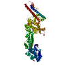
| Structure viewer | Molecule: MolmilJmol/JSmol |
|---|
- Downloads & links
-Download
| PDBx/mmCIF format | 2hft.cif.gz | 59.4 KB | Display | PDBx/mmCIF format |
|---|---|---|---|---|
| PDB format | pdb2hft.ent.gz | 43.4 KB | Display | PDB format |
| PDBx/mmJSON format | 2hft.json.gz | Tree view | PDBx/mmJSON format | |
| Others |  Other downloads Other downloads |
-Validation report
| Arichive directory | https://data.pdbj.org/pub/pdb/validation_reports/hf/2hftftp://data.pdbj.org/pub/pdb/validation_reports/hf/2hft | HTTPS FTP |
|---|
-Related structure data
| Similar structure data |
|---|
-Links
 PDBj
PDBj


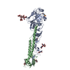
- Assembly
| Deposited unit | 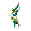
| ||||||||
|---|---|---|---|---|---|---|---|---|---|
| 1 | 
| ||||||||
| Unit cell |
| ||||||||
| Atom site foot note | 1: CIS PROLINE - PRO 27 |
-Components
| #1: Protein | Mass: 24697.398 Da / Num. of mol.: 1 Source method: isolated from a genetically manipulated source Source: (gene. exp.) Homo sapiens (human) / Production host:  |
|---|---|
| #2: Chemical | ChemComp-SO4 /   Mass: 96.063 Da / Num. of mol.: 1 / Source method: obtained synthetically / Formula: SO4 Mass: 96.063 Da / Num. of mol.: 1 / Source method: obtained synthetically / Formula: SO4 |
| #3: Water | ChemComp-HOH /  Mass: 18.015 Da / Num. of mol.: 235 / Source method: isolated from a natural source / Formula: H2O Mass: 18.015 Da / Num. of mol.: 235 / Source method: isolated from a natural source / Formula: H2O |
| Compound details | THE FACTOR VII BINDING SITE WAS IDENTIFIED BY ALA-SCANNING MUTAGENESIS (KELLEY ET AL., 1995; SEE ...THE FACTOR VII BINDING SITE WAS IDENTIFIED |
| Has protein modification | Y |
-Experimental details
-Experiment
| Experiment | Method: X-RAY DIFFRACTION / Number of used crystals: 1 |
|---|
- Sample preparation
| Crystal | Density Matthews: 2.39 Å3/Da / Density % sol: 48.44 % | |||||||||||||||||||||||||||||||||||
|---|---|---|---|---|---|---|---|---|---|---|---|---|---|---|---|---|---|---|---|---|---|---|---|---|---|---|---|---|---|---|---|---|---|---|---|---|
| Crystal grow | pH: 7.5 / Details: pH 7.5 | |||||||||||||||||||||||||||||||||||
| Crystal | *PLUS Density % sol: 49 % | |||||||||||||||||||||||||||||||||||
| Crystal grow | *PLUS Temperature: 100 K / Method: vapor diffusion, sitting dropDetails: drop contains 0.05 ml of protein solution and 0.01 ml of reservoir solution | |||||||||||||||||||||||||||||||||||
| Components of the solutions | *PLUS
|
-Data collection
| Diffraction | Mean temperature: 100 K |
|---|---|
| Diffraction source | Wavelength: 0.908 Å |
| Detector | Type: FUJI FILM / Detector: IMAGE PLATE |
| Radiation | Scattering type: x-ray |
| Radiation wavelength | Wavelength: 0.908 Å / Relative weight: 1 |
| Reflection | Resolution: 1.69→6 Å / Num. obs: 24199 / % possible obs: 97 % / Redundancy: 4.5 % / Rmerge(I) obs: 0.058 |
| Reflection | *PLUS Rmerge(I) obs: 0.058 |
- Processing
| Software |
| |||||||||||||||||||||||||||||||||||||||||||||||||||||||||||||||
|---|---|---|---|---|---|---|---|---|---|---|---|---|---|---|---|---|---|---|---|---|---|---|---|---|---|---|---|---|---|---|---|---|---|---|---|---|---|---|---|---|---|---|---|---|---|---|---|---|---|---|---|---|---|---|---|---|---|---|---|---|---|---|---|---|
| Refinement | Resolution: 1.69→6 Å / σ(F): 0
| |||||||||||||||||||||||||||||||||||||||||||||||||||||||||||||||
| Displacement parameters | Biso mean: 27.6 Å2 | |||||||||||||||||||||||||||||||||||||||||||||||||||||||||||||||
| Refine analyze | Luzzati coordinate error obs: 0.2 Å | |||||||||||||||||||||||||||||||||||||||||||||||||||||||||||||||
| Refinement step | Cycle: LAST / Resolution: 1.69→6 Å
| |||||||||||||||||||||||||||||||||||||||||||||||||||||||||||||||
| Refine LS restraints |
| |||||||||||||||||||||||||||||||||||||||||||||||||||||||||||||||
| Software | *PLUS Name: PROLSQ/X-PLOR / Classification: refinement | |||||||||||||||||||||||||||||||||||||||||||||||||||||||||||||||
| Refine LS restraints | *PLUS
|
