 Movie
Movie Controller

[English] 日本語
 Yorodumi
- PDB-5g51: High resolution structure of the part of VP3 protein of Deformed ... -
+ Open data

- Basic information
| Entry | Database: PDB / ID: 5g51 | |||||||||
|---|---|---|---|---|---|---|---|---|---|---|
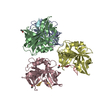
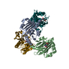
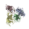
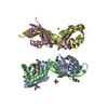
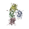
| Title | High resolution structure of the part of VP3 protein of Deformed Wing Virus forming P-domain | |||||||||
 Components Components | DWV-VP3-P-DOMAIN | |||||||||
 Keywords Keywords | VIRAL PROTEIN / PICORNAVIRALES / PICORNAVIRALES IFLAVIRIDAE IFLAVIRUS DWV CAPSID P-DOMAIN JELLYROLL INHIBITOR ANTIVIRAL CATALYTIC SITE PROTEASE LIPASE ESTERASE RECEPTOR | |||||||||
| Function / homology |  Function and homology information Function and homology informationviral capsid / Hydrolases; Acting on peptide bonds (peptidases); Cysteine endopeptidases / RNA helicase activity / RNA helicase / RNA-directed RNA polymerase / cysteine-type endopeptidase activity / viral RNA genome replication / RNA-directed RNA polymerase activity / structural molecule activity / DNA-templated transcription ...viral capsid / Hydrolases; Acting on peptide bonds (peptidases); Cysteine endopeptidases / RNA helicase activity / RNA helicase / RNA-directed RNA polymerase / cysteine-type endopeptidase activity / viral RNA genome replication / RNA-directed RNA polymerase activity / structural molecule activity / DNA-templated transcription / proteolysis / RNA binding / ATP binding Similarity search - Function | |||||||||
| Biological species |  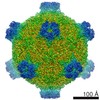 DEFORMED WING VIRUS DEFORMED WING VIRUS | |||||||||
| Method |  X-RAY DIFFRACTION / SYNCHROTRON / MOLECULAR REPLACEMENT / Resolution: 1.45 Å X-RAY DIFFRACTION / SYNCHROTRON / MOLECULAR REPLACEMENT / Resolution: 1.45 Å | |||||||||
 Authors Authors | Skubnik, K. / Novacek, J. / Fuzik, T. / Pridal, A. / Paxton, R. / Plevka, P. | |||||||||
 Citation Citation | Journal: Proc Natl Acad Sci U S A / Year: 2017 Title: Structure of deformed wing virus, a major honey bee pathogen. Authors: Karel Škubník / Jiří Nováček / Tibor Füzik / Antonín Přidal / Robert J Paxton / Pavel Plevka /   Abstract: The worldwide population of western honey bees () is under pressure from habitat loss, environmental stress, and pathogens, particularly viruses that cause lethal epidemics. Deformed wing virus (DWV) ...The worldwide population of western honey bees () is under pressure from habitat loss, environmental stress, and pathogens, particularly viruses that cause lethal epidemics. Deformed wing virus (DWV) from the family , together with its vector, the mite , is likely the major threat to the world's honey bees. However, lack of knowledge of the atomic structures of iflaviruses has hindered the development of effective treatments against them. Here, we present the virion structures of DWV determined to a resolution of 3.1 Å using cryo-electron microscopy and 3.8 Å by X-ray crystallography. The C-terminal extension of capsid protein VP3 folds into a globular protruding (P) domain, exposed on the virion surface. The P domain contains an Asp-His-Ser catalytic triad that is, together with five residues that are spatially close, conserved among iflaviruses. These residues may participate in receptor binding or provide the protease, lipase, or esterase activity required for entry of the virus into a host cell. Furthermore, nucleotides of the DWV RNA genome interact with VP3 subunits. The capsid protein residues involved in the RNA binding are conserved among honey bee iflaviruses, suggesting a putative role of the genome in stabilizing the virion or facilitating capsid assembly. Identifying the RNA-binding and putative catalytic sites within the DWV virion structure enables future analyses of how DWV and other iflaviruses infect insect cells and also opens up possibilities for the development of antiviral treatments. | |||||||||
| History |
|
- Structure visualization
| Structure viewer | Molecule: MolmilJmol/JSmol |
|---|
- Downloads & links
-Download
| PDBx/mmCIF format | 5g51.cif.gz | 53.2 KB | Display | PDBx/mmCIF format |
|---|---|---|---|---|
| PDB format | pdb5g51.ent.gz | 39.2 KB | Display | PDB format |
| PDBx/mmJSON format | 5g51.json.gz | Tree view | PDBx/mmJSON format | |
| Others |  Other downloads Other downloads |
-Validation report
| Arichive directory | https://data.pdbj.org/pub/pdb/validation_reports/g5/5g51ftp://data.pdbj.org/pub/pdb/validation_reports/g5/5g51 | HTTPS FTP |
|---|
-Related structure data
| Related structure data | 3570C 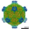 3574C 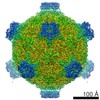 3575C 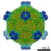 4009C 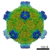 4014C 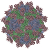 5g52C  5l7qC  5l8qC 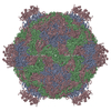 5mupC 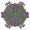 5mv5C 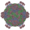 5mv6C C: citing same article ( |
|---|---|
| Similar structure data |


-Links
 PDBj
PDBj
- Assembly
| Deposited unit | 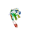
| |||||||||||||||
|---|---|---|---|---|---|---|---|---|---|---|---|---|---|---|---|---|
| 1 | 
| |||||||||||||||
| Unit cell |
| |||||||||||||||
| Components on special symmetry positions |
|
-Components
| #1: Protein | Mass: 17535.582 Da / Num. of mol.: 1 / Fragment: P-DOMAIN, RESIDUES 260-398 Source method: isolated from a genetically manipulated source Details: PART OF THE VP3 CHAIN OF THE DEFORMED WING VIRUS. / Source: (gene. exp.) DEFORMED WING VIRUS / Plasmid: PET22T / Production host:  |
|---|---|
| #2: Water | ChemComp-HOH /  Mass: 18.015 Da / Num. of mol.: 242 / Source method: isolated from a natural source / Formula: H2O Mass: 18.015 Da / Num. of mol.: 242 / Source method: isolated from a natural source / Formula: H2O |
| Sequence details | SEQUENCE WILL BE UPLOAD SOON TO THE UNIPROT DATABASE. |
-Experimental details
-Experiment
| Experiment | Method: X-RAY DIFFRACTION / Number of used crystals: 1 |
|---|
- Sample preparation
| Crystal | Density Matthews: 3.16 Å3/Da / Density % sol: 61.09 % / Description: NONE |
|---|---|
| Crystal grow | pH: 7.5 / Details: 0.1 M HEPES PH 7.5, 4.3 M NACL, |
-Data collection
| Diffraction | Mean temperature: 100 K |
|---|---|
| Diffraction source | Source: SYNCHROTRON / Site: SOLEIL  / Beamline: PROXIMA 1 / Wavelength: 0.97857 / Beamline: PROXIMA 1 / Wavelength: 0.97857 |
| Detector | Type: DECTRIS PILATUS 6M / Detector: PIXEL / Date: Mar 24, 2016 Details: KIRKPATRICK-BAEZ PAIR OF BI-MORPH MIRRORS PLUS CHANNEL |
| Radiation | Monochromator: CRYOGENICALLY COOLED MONOCHROMATOR CRYSTAL / Protocol: SINGLE WAVELENGTH / Monochromatic (M) / Laue (L): M / Scattering type: x-ray |
| Radiation wavelength | Wavelength: 0.97857 Å / Relative weight: 1 |
| Reflection | Resolution: 1.45→39.75 Å / Num. obs: 34920 / % possible obs: 99.9 % / Observed criterion σ(I): 2.1 / Redundancy: 6.09 % / Rmerge(I) obs: 0.06 / Net I/σ(I): 14.2 |
| Reflection shell | Resolution: 1.45→1.47 Å / Redundancy: 5.36 % / Rmerge(I) obs: 0.54 / Mean I/σ(I) obs: 2.1 / % possible all: 98.4 |
- Processing
| Software |
| ||||||||||||||||||||||||||||||||||||||||||||||||||||||||||||||||||||||||||||||||||||||||||||||||||
|---|---|---|---|---|---|---|---|---|---|---|---|---|---|---|---|---|---|---|---|---|---|---|---|---|---|---|---|---|---|---|---|---|---|---|---|---|---|---|---|---|---|---|---|---|---|---|---|---|---|---|---|---|---|---|---|---|---|---|---|---|---|---|---|---|---|---|---|---|---|---|---|---|---|---|---|---|---|---|---|---|---|---|---|---|---|---|---|---|---|---|---|---|---|---|---|---|---|---|---|
| Refinement | Method to determine structure: MOLECULAR REPLACEMENT / Resolution: 1.45→39.749 Å / SU ML: 0.17 / σ(F): 1.39 / Phase error: 22.09 / Stereochemistry target values: ML
| ||||||||||||||||||||||||||||||||||||||||||||||||||||||||||||||||||||||||||||||||||||||||||||||||||
| Solvent computation | Shrinkage radii: 0.9 Å / VDW probe radii: 1.11 Å / Solvent model: FLAT BULK SOLVENT MODEL | ||||||||||||||||||||||||||||||||||||||||||||||||||||||||||||||||||||||||||||||||||||||||||||||||||
| Refinement step | Cycle: LAST / Resolution: 1.45→39.749 Å
| ||||||||||||||||||||||||||||||||||||||||||||||||||||||||||||||||||||||||||||||||||||||||||||||||||
| Refine LS restraints |
| ||||||||||||||||||||||||||||||||||||||||||||||||||||||||||||||||||||||||||||||||||||||||||||||||||
| LS refinement shell |
|
